Copyright
©2013 Baishideng Publishing Group Co.
World J Clin Pediatr. Nov 8, 2013; 2(4): 54-64
Published online Nov 8, 2013. doi: 10.5409/wjcp.v2.i4.54
Published online Nov 8, 2013. doi: 10.5409/wjcp.v2.i4.54
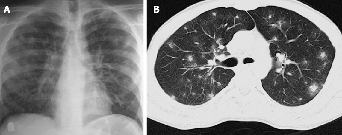
Figure 5 A 17-year-old boy with acute lymphocytic leukemia along with angioinvasive aspergillosis.
Chest radiograph (A) showing multiple fluffy nodules in bilateral lung fields. High resolution CT image (B) of the same patient shows multiple nodules with surrounding ground glass opacity (halo sign).
- Citation: Singh D, Bhalla AS, Veedu PT, Arora A. Imaging evaluation of hemoptysis in children. World J Clin Pediatr 2013; 2(4): 54-64
- URL: https://www.wjgnet.com/2219-2808/full/v2/i4/54.htm
- DOI: https://dx.doi.org/10.5409/wjcp.v2.i4.54


