Copyright
©2013 Baishideng Publishing Group Co.
World J Clin Pediatr. Nov 8, 2013; 2(4): 36-45
Published online Nov 8, 2013. doi: 10.5409/wjcp.v2.i4.36
Published online Nov 8, 2013. doi: 10.5409/wjcp.v2.i4.36
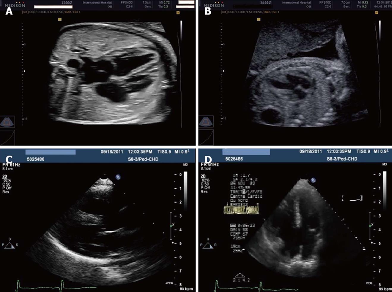
Figure 1 Ultrasonography.
A: Normal 4-chamber view by fetal echocardiography at 26 wk gestation; B: Four-chamber view by fetal echocardiography at 22 wk gestation showing ventricular septal defects; C: Left-parasternal long-axis view in an infant with Down syndrome and complete atrioventricular (AV) canal and pulmonary hypertension; D: Apical 4-chamber view in an infant with Down syndrome and complete AV canal and pulmonary hypertension.
- Citation: Al-Biltagi MA. Echocardiography in children with Down syndrome. World J Clin Pediatr 2013; 2(4): 36-45
- URL: https://www.wjgnet.com/2219-2808/full/v2/i4/36.htm
- DOI: https://dx.doi.org/10.5409/wjcp.v2.i4.36


