Copyright
©2013 Baishideng Publishing Group Co.
World J Clin Pediatr. Aug 8, 2013; 2(3): 26-30
Published online Aug 8, 2013. doi: 10.5409/wjcp.v2.i3.26
Published online Aug 8, 2013. doi: 10.5409/wjcp.v2.i3.26
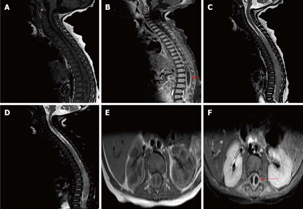
Figure 2 Magnetic resonance imaging spine.
An intra-spinal and extra-dural cystic lesion of low signal intensity non-contrast sagittal T1-weighted imaging (T1WI) (A), non-contrast axial T1WI (E). It shows an intense marginal enhancement post-contrast sagittal T1WI (B, red arrow) and post-contrast axial T1WI (F, red arrow). The lesion extends up to the cervical spine sagittal T2WI (C) and lateral to the left side of the spinal canal sagittal T2WI (D).
- Citation: Mostafa M, Nasef N, Barakat T, El-Hawary AK, Abdel-Hady H. Acute flaccid paralysis in a patient with sacral dimple. World J Clin Pediatr 2013; 2(3): 26-30
- URL: https://www.wjgnet.com/2219-2808/full/v2/i3/26.htm
- DOI: https://dx.doi.org/10.5409/wjcp.v2.i3.26


