Copyright
©The Author(s) 2025.
World J Clin Pediatr. Jun 9, 2025; 14(2): 101080
Published online Jun 9, 2025. doi: 10.5409/wjcp.v14.i2.101080
Published online Jun 9, 2025. doi: 10.5409/wjcp.v14.i2.101080
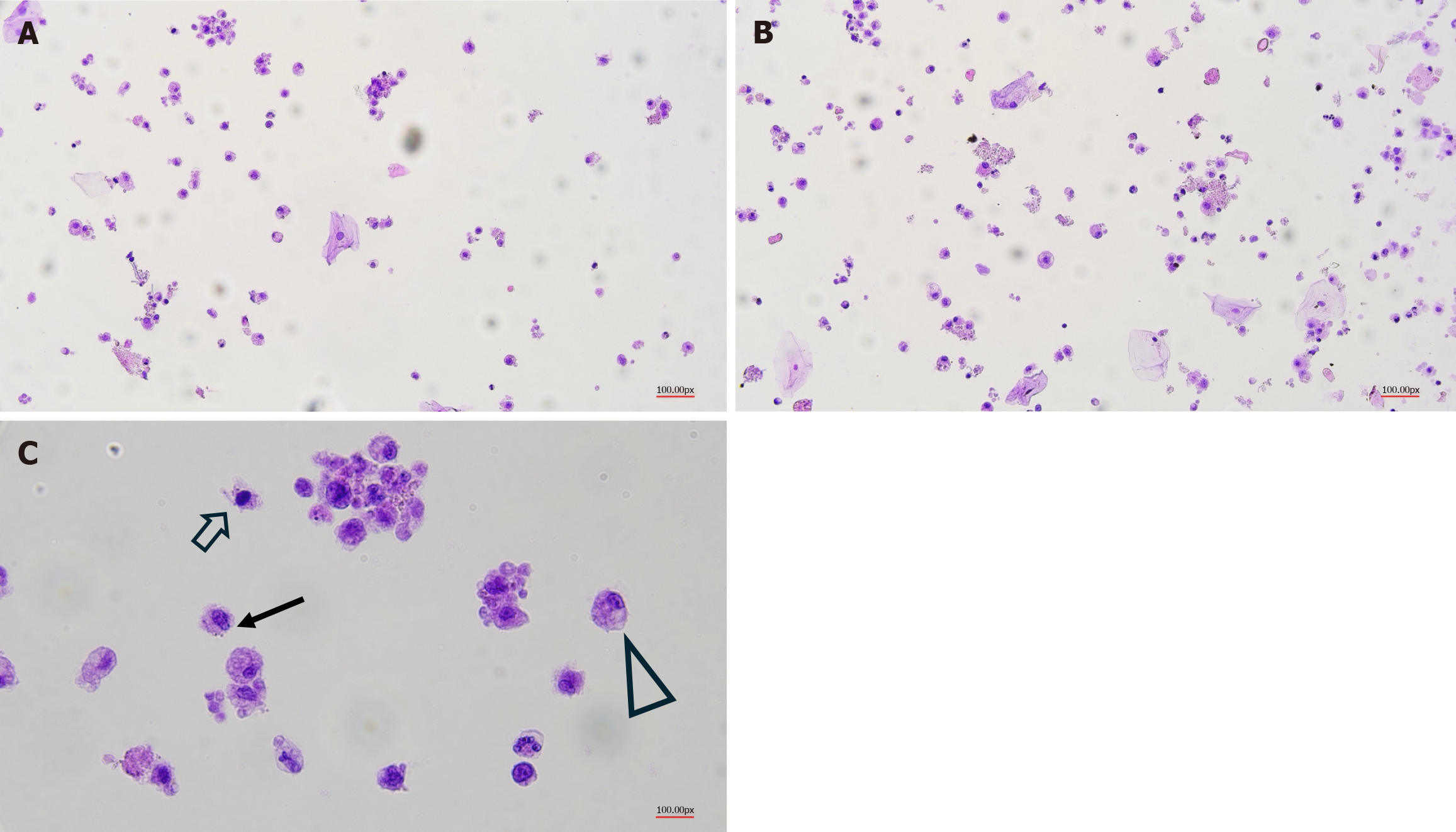
Figure 1 Different example of cells isolated with from fresh human milk.
A-C: These cells, fixed and stained using our cytological methods routinely used in our laboratories, are characterized by different morphological aspects, underling the presence of a heterogeneous cell population. Briefly, breast milk samples were recruited from six healthy volunteers. The experimental analysis were authorized by the Comitato Etico Indipendente Ospedaliero (Università di Cagliari; Prot. PG/2022/795). Breast milk cells were isolated from fresh milk, diluted with an equal volume of Dulbecco’s phosphate-buffered saline (pH 7.4; Gibco) and centrifugated at 810 g for 20 minutes at 20 °C. The fat layer and liquid were removed, while the cell pellet was washed twice with Dulbecco’s phosphate-buffered saline. The pellet was fixed with ThinPrep solution (Hologic, Inc. www.hologic.com) and the cell samples were processed for immunocytochemistry analysis with a ThinPrep 5000 Processor. Different arrows highlight possible different cell types. Solid arrow: Stem cells; Hollow arrow: Immune cells; Triangle: Milk-secretory cells.
- Citation: Faa G, Pichiri G, Coni P, Dessì A, Fraschini M, Fanos V. They will be famous: Multipotent stem cells in breast milk. World J Clin Pediatr 2025; 14(2): 101080
- URL: https://www.wjgnet.com/2219-2808/full/v14/i2/101080.htm
- DOI: https://dx.doi.org/10.5409/wjcp.v14.i2.101080


