Copyright
©The Author(s) 2025.
World J Clin Pediatr. Mar 9, 2025; 14(1): 99288
Published online Mar 9, 2025. doi: 10.5409/wjcp.v14.i1.99288
Published online Mar 9, 2025. doi: 10.5409/wjcp.v14.i1.99288
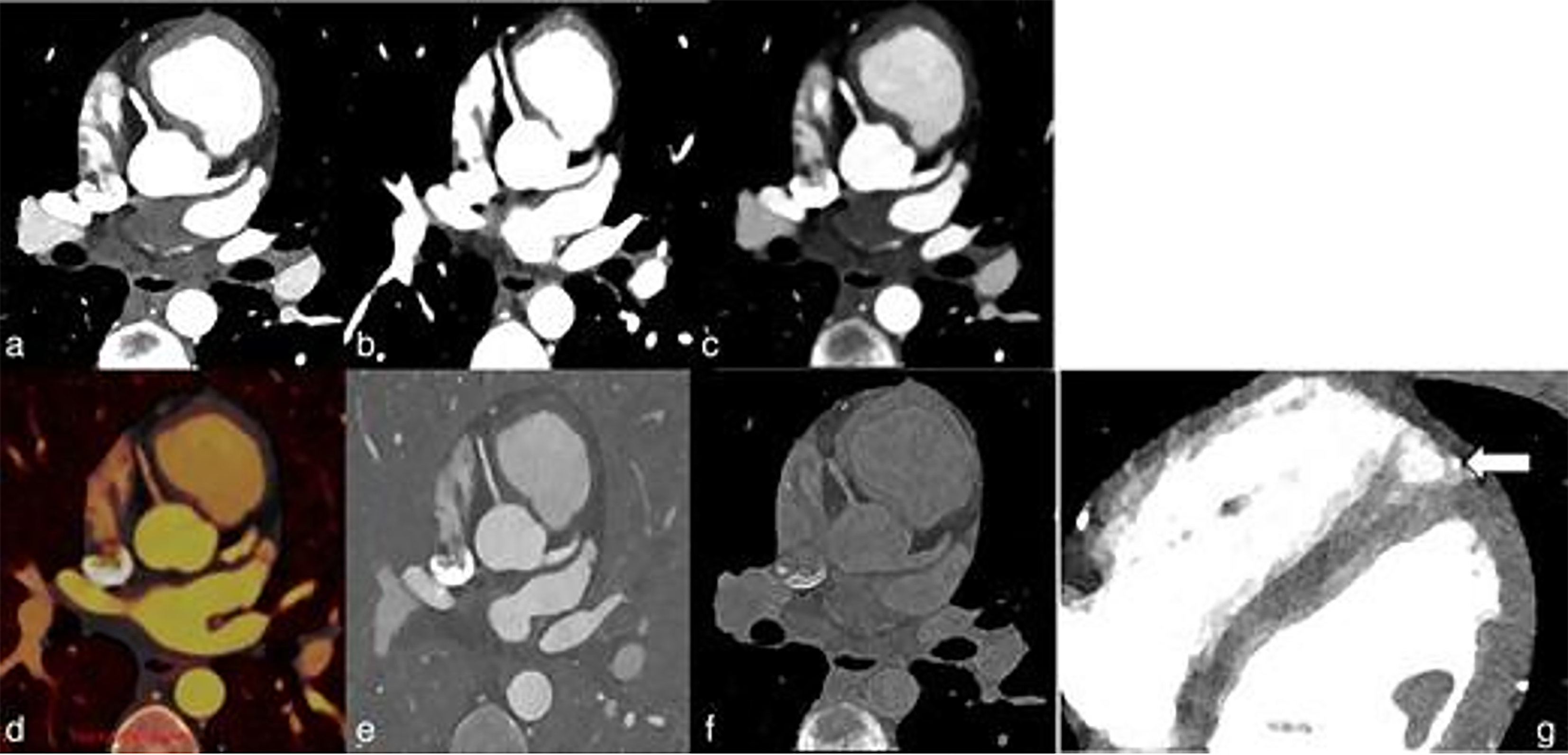
Figure 3 A 16-year-old male with a suspected coronary anomaly.
a: Cardiac multienergy photon-counting detectors (PCDs) scan reconstructions at 40 keV; b: Cardiac multienergy PCDs scan reconstructions at 50 keV; c: Cardiac multienergy PCDs scan reconstructions at 100 keV; d: Cardiac multienergy PCDs scan reconstructions at color-coded iodine overlay; e: Cardiac multienergy PCDs scan reconstructions at iodine overlay; f: Cardiac multienergy PCDs scan reconstructions at virtual non-contrast; g: No coronary anomaly was detected but a short segment myocardial bridge (arrow) of the distal left anterior descending artery at the level of the apex was detected as shown in image. Copyright ©Siemens Healthineers AG 2024. See: https://www.siemens-healthineers.com/press/copyright#:~:text=Press%20materials%3A%20Copyright&text=Materials%20used%20for%20editorial%20purposes,electronically%20manipulated%20form%2C%20is%20prohibited.
- Citation: Perera Molligoda Arachchige AS, Verma Y. Role of photon-counting computed tomography in pediatric cardiovascular imaging. World J Clin Pediatr 2025; 14(1): 99288
- URL: https://www.wjgnet.com/2219-2808/full/v14/i1/99288.htm
- DOI: https://dx.doi.org/10.5409/wjcp.v14.i1.99288


