Copyright
©The Author(s) 2025.
World J Clin Pediatr. Mar 9, 2025; 14(1): 99177
Published online Mar 9, 2025. doi: 10.5409/wjcp.v14.i1.99177
Published online Mar 9, 2025. doi: 10.5409/wjcp.v14.i1.99177
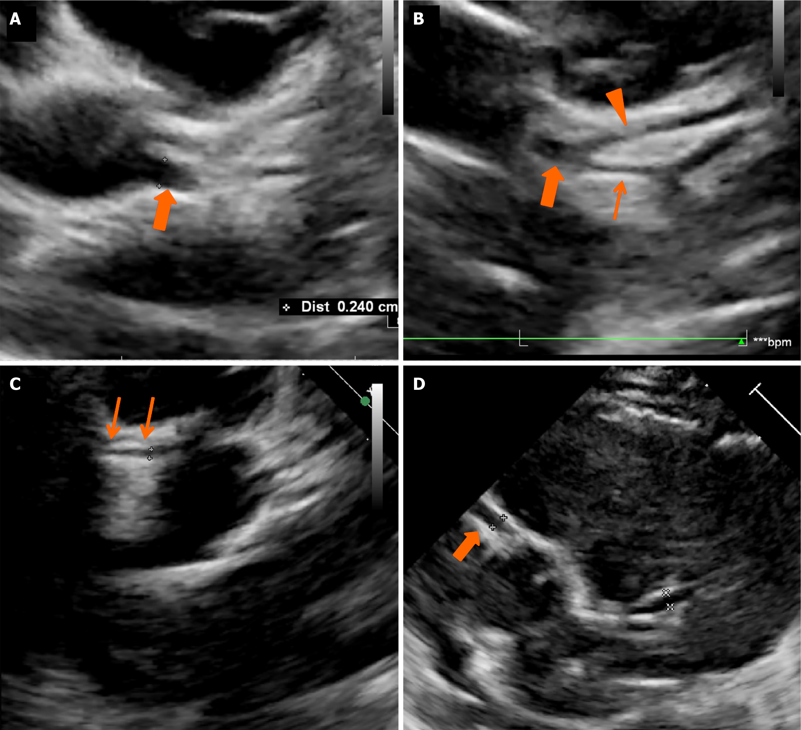
Figure 2 2D echocardiography images in patient 2 and patient 3.
A-C: Images show dilated left main coronary artery (thick arrows in A and B) with normal left anterior descending (arrowhead) and left circumflex (thin arrow in B) and normal proximal right coronary artery (thin arrows in C); D: Image shows dilated proximal right coronary artery in patient 3 (thick arrow).
- Citation: Pilania RK, Nadig PL, Basu S, Tyagi R, Thangaraj A, Aggarwal R, Arora M, Sharma A, Singh S, Singhal M. Congenital anomalies of coronary artery misdiagnosed as coronary dilatations in Kawasaki disease: A clinical predicament. World J Clin Pediatr 2025; 14(1): 99177
- URL: https://www.wjgnet.com/2219-2808/full/v14/i1/99177.htm
- DOI: https://dx.doi.org/10.5409/wjcp.v14.i1.99177


