Copyright
©The Author(s) 2023.
World J Clin Pediatr. Jun 9, 2023; 12(3): 97-106
Published online Jun 9, 2023. doi: 10.5409/wjcp.v12.i3.97
Published online Jun 9, 2023. doi: 10.5409/wjcp.v12.i3.97
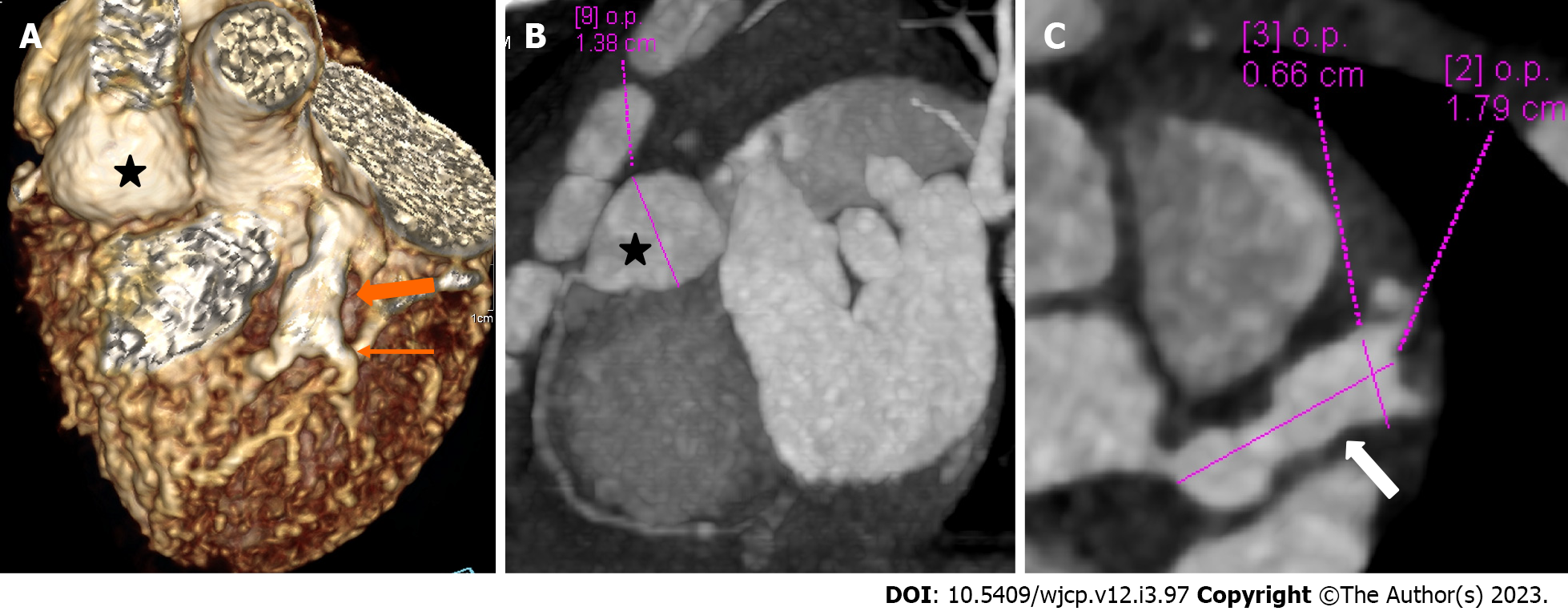
Figure 4 Panel of computed tomography coronary angiography images showing its ability to precisely identify location and morphology of aneurysms with extension into side branches.
Computed tomography coronary angiography (A: Volume rendered image; B: Curved reformatted image and C-axial image) of 4 years female child in acute phase (presentation) demonstrate giant saccular aneurysm in proximal resonance coronary angiography (asterisk in A and B). Fusiform aneurysm is seen in proximal left anterior descending (thick arrow in A) with extension into diagonal-1 branch (thin arrow in A).
- Citation: Singhal M, Pilania RK, Gupta P, Johnson N, Singh S. Emerging role of computed tomography coronary angiography in evaluation of children with Kawasaki disease. World J Clin Pediatr 2023; 12(3): 97-106
- URL: https://www.wjgnet.com/2219-2808/full/v12/i3/97.htm
- DOI: https://dx.doi.org/10.5409/wjcp.v12.i3.97


