Copyright
©The Author(s) 2023.
World J Clin Pediatr. Jun 9, 2023; 12(3): 97-106
Published online Jun 9, 2023. doi: 10.5409/wjcp.v12.i3.97
Published online Jun 9, 2023. doi: 10.5409/wjcp.v12.i3.97
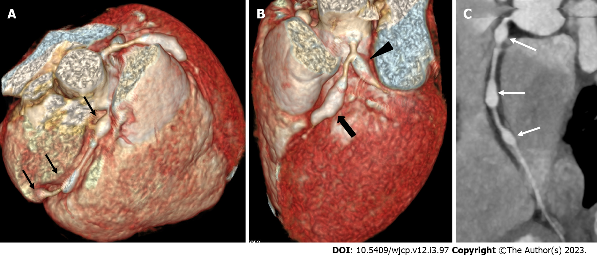
Figure 3 Computed tomography coronary angiography images showing its ability to evaluate mid and distal segments of coronary arteries.
Computed tomography coronary angiography (A and B: Volume rendered images; C: Curved reformatted image of RCA) of 4 years male at presentation demonstrate skip fusiform aneurysms in RCA (Thin arrows in A and C) and fusiform aneurysms in proximal LAD (thick arrow in B) and proximal left circumflex (arrow head in B). Fusiform aneurysm in mid and distal RCA could not be visualized on transthoracic echocardiography.
- Citation: Singhal M, Pilania RK, Gupta P, Johnson N, Singh S. Emerging role of computed tomography coronary angiography in evaluation of children with Kawasaki disease. World J Clin Pediatr 2023; 12(3): 97-106
- URL: https://www.wjgnet.com/2219-2808/full/v12/i3/97.htm
- DOI: https://dx.doi.org/10.5409/wjcp.v12.i3.97


