Copyright
©The Author(s) 2022.
World J Clin Pediatr. May 9, 2022; 11(3): 270-288
Published online May 9, 2022. doi: 10.5409/wjcp.v11.i3.270
Published online May 9, 2022. doi: 10.5409/wjcp.v11.i3.270
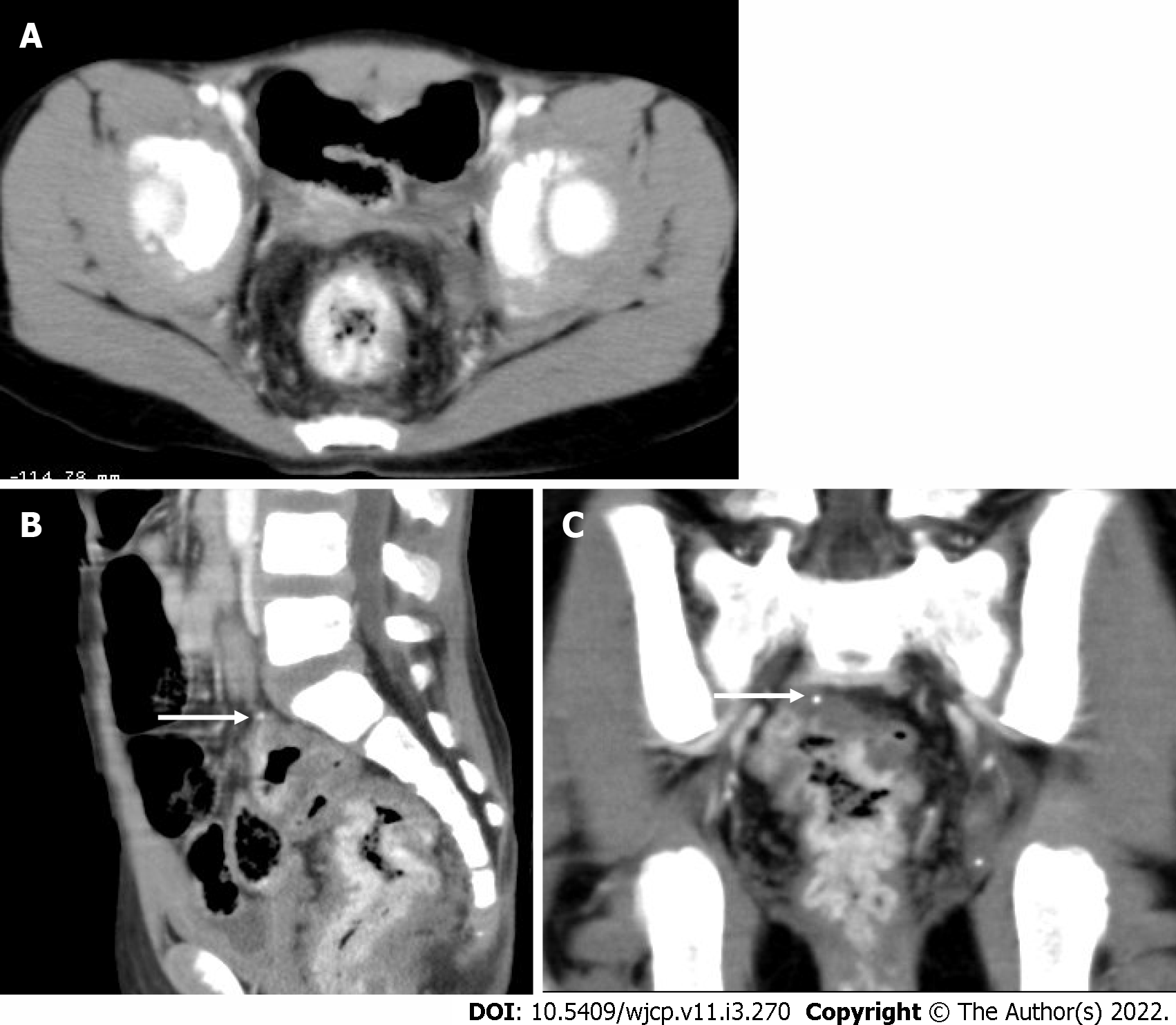
Figure 17 An 8-year-old girl with bleeding per rectum.
Axial (A), sagittal (B), and coronal (C) contrast enhanced computed tomography images showing diffuse rectal wall thickening with intense contrast enhancement. A tiny phlebolith is also seen (arrows in B and C). Findings are suggestive of rectal hemangioma.
- Citation: Chandel K, Jain R, Bhatia A, Saxena AK, Sodhi KS. Bleeding per rectum in pediatric population: A pictorial review. World J Clin Pediatr 2022; 11(3): 270-288
- URL: https://www.wjgnet.com/2219-2808/full/v11/i3/270.htm
- DOI: https://dx.doi.org/10.5409/wjcp.v11.i3.270


