Copyright
©The Author(s) 2022.
World J Clin Pediatr. May 9, 2022; 11(3): 270-288
Published online May 9, 2022. doi: 10.5409/wjcp.v11.i3.270
Published online May 9, 2022. doi: 10.5409/wjcp.v11.i3.270
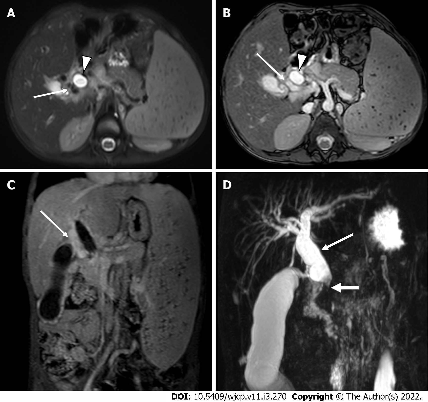
Figure 14 Portal cavernoma cholangiopathy.
A and B: Axial T2 weighted and axial BTFE magnetic resonance images show dilated CBD [arrowhead] with multiple pericholedochal collaterals [white arrow]; C: Coronal post-contrast T1 image shows multiple enhancing pericholedochal collaterals [white arrow]; D: Massive splenomegaly. Thick slab coronal magnetic resonance cholangiopancreatography image shows dilated and tortuous CBD (thin arrow) with abrupt cut-off at the lower end (thick arrow).
- Citation: Chandel K, Jain R, Bhatia A, Saxena AK, Sodhi KS. Bleeding per rectum in pediatric population: A pictorial review. World J Clin Pediatr 2022; 11(3): 270-288
- URL: https://www.wjgnet.com/2219-2808/full/v11/i3/270.htm
- DOI: https://dx.doi.org/10.5409/wjcp.v11.i3.270


