Copyright
©The Author(s) 2022.
World J Clin Pediatr. May 9, 2022; 11(3): 270-288
Published online May 9, 2022. doi: 10.5409/wjcp.v11.i3.270
Published online May 9, 2022. doi: 10.5409/wjcp.v11.i3.270
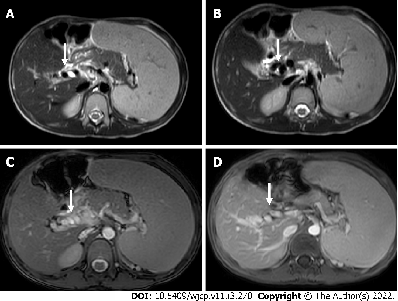
Figure 13 A 7-year-old boy with extra-hepatic portal vein obstruction.
A and B: Axial T2W images showing non visualized main portal vein with multiple collaterals (arrow) along the course of the portal vein; C and D: Axial BTFE and post-contrast T1 images show multiple collaterals (arrow) replacing the main portal vein at the porta. In addition, splenomegaly is also seen.
- Citation: Chandel K, Jain R, Bhatia A, Saxena AK, Sodhi KS. Bleeding per rectum in pediatric population: A pictorial review. World J Clin Pediatr 2022; 11(3): 270-288
- URL: https://www.wjgnet.com/2219-2808/full/v11/i3/270.htm
- DOI: https://dx.doi.org/10.5409/wjcp.v11.i3.270


