Copyright
©The Author(s) 2022.
World J Clin Pediatr. May 9, 2022; 11(3): 270-288
Published online May 9, 2022. doi: 10.5409/wjcp.v11.i3.270
Published online May 9, 2022. doi: 10.5409/wjcp.v11.i3.270
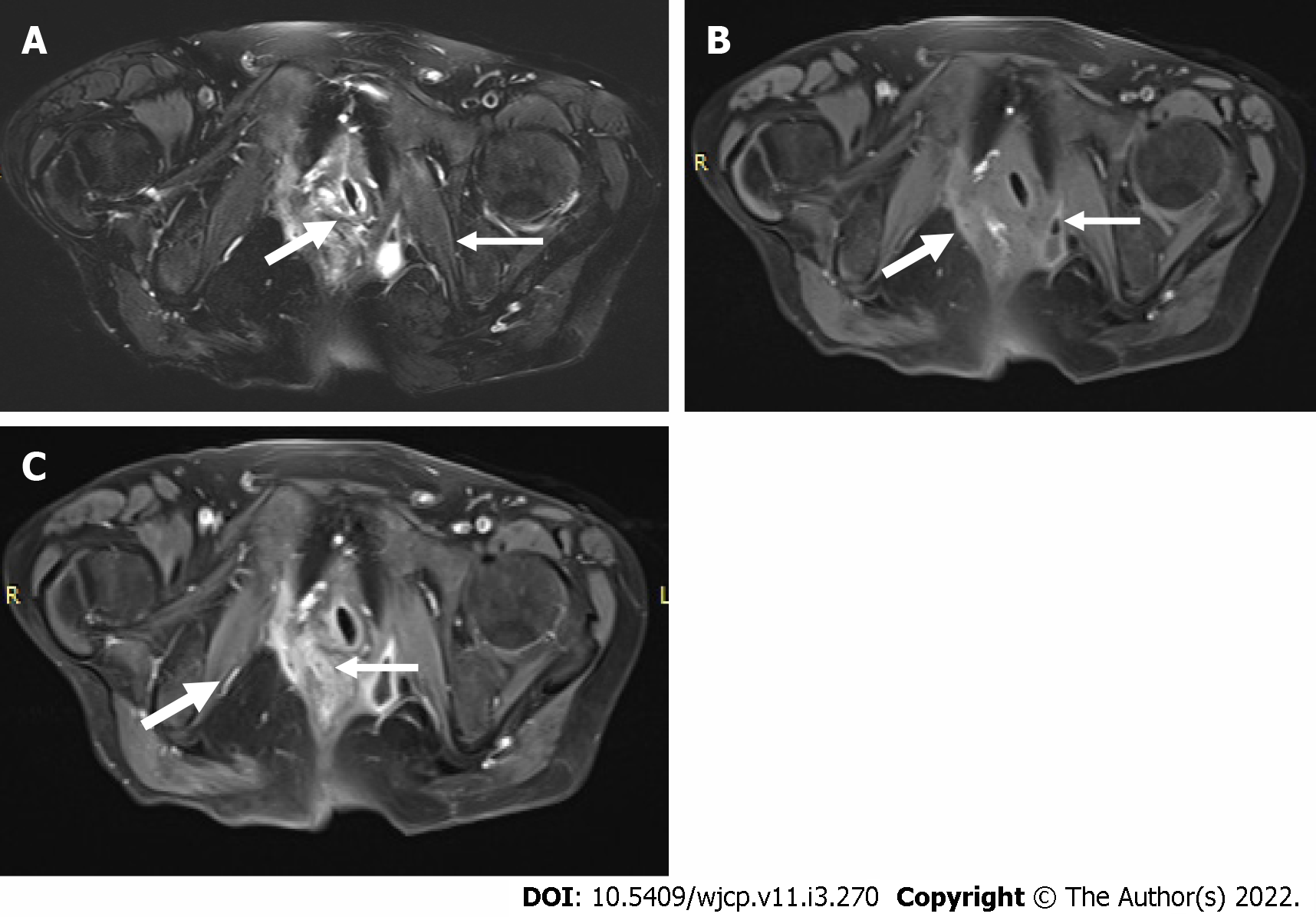
Figure 8 Magnetic resonance image pelvis images in a child with Crohn’s disease.
A and B: T2 weighted fat suppressed and pre contrast T1 weighted images showing a fistulous tract (thick arrow in A and B) on the right side communicating with the rectum in the midline. In addition, a small collection (thin arrow in A and B) with air focus is seen in the left ischio-rectal fossa; C: Post contrast T1 weighted fat suppressed image showing enhancement of the tract (thick arrow) as well as peripheral enhancement of the collection (thin arrow).
- Citation: Chandel K, Jain R, Bhatia A, Saxena AK, Sodhi KS. Bleeding per rectum in pediatric population: A pictorial review. World J Clin Pediatr 2022; 11(3): 270-288
- URL: https://www.wjgnet.com/2219-2808/full/v11/i3/270.htm
- DOI: https://dx.doi.org/10.5409/wjcp.v11.i3.270


