Copyright
©The Author(s) 2022.
World J Clin Pediatr. May 9, 2022; 11(3): 270-288
Published online May 9, 2022. doi: 10.5409/wjcp.v11.i3.270
Published online May 9, 2022. doi: 10.5409/wjcp.v11.i3.270
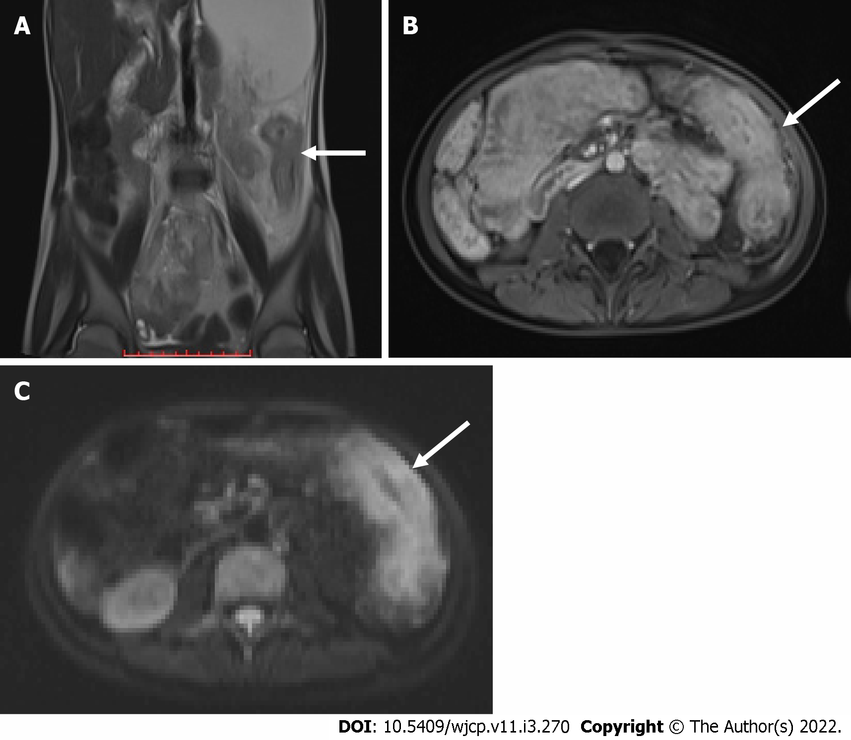
Figure 7 Diffusion weighted imaging.
A: Coronal T2 weighted magnetic resonance image showing long segment mural thickening in the descending colon (arrow in A) which is showing post contrast enhancement in post contrast T1 image (arrow in B) and intense diffusion restriction in diffusion weighted image (arrow in C) suggestive of active disease.
- Citation: Chandel K, Jain R, Bhatia A, Saxena AK, Sodhi KS. Bleeding per rectum in pediatric population: A pictorial review. World J Clin Pediatr 2022; 11(3): 270-288
- URL: https://www.wjgnet.com/2219-2808/full/v11/i3/270.htm
- DOI: https://dx.doi.org/10.5409/wjcp.v11.i3.270


