Copyright
©The Author(s) 2021.
World J Clin Pediatr. Nov 9, 2021; 10(6): 124-136
Published online Nov 9, 2021. doi: 10.5409/wjcp.v10.i6.124
Published online Nov 9, 2021. doi: 10.5409/wjcp.v10.i6.124
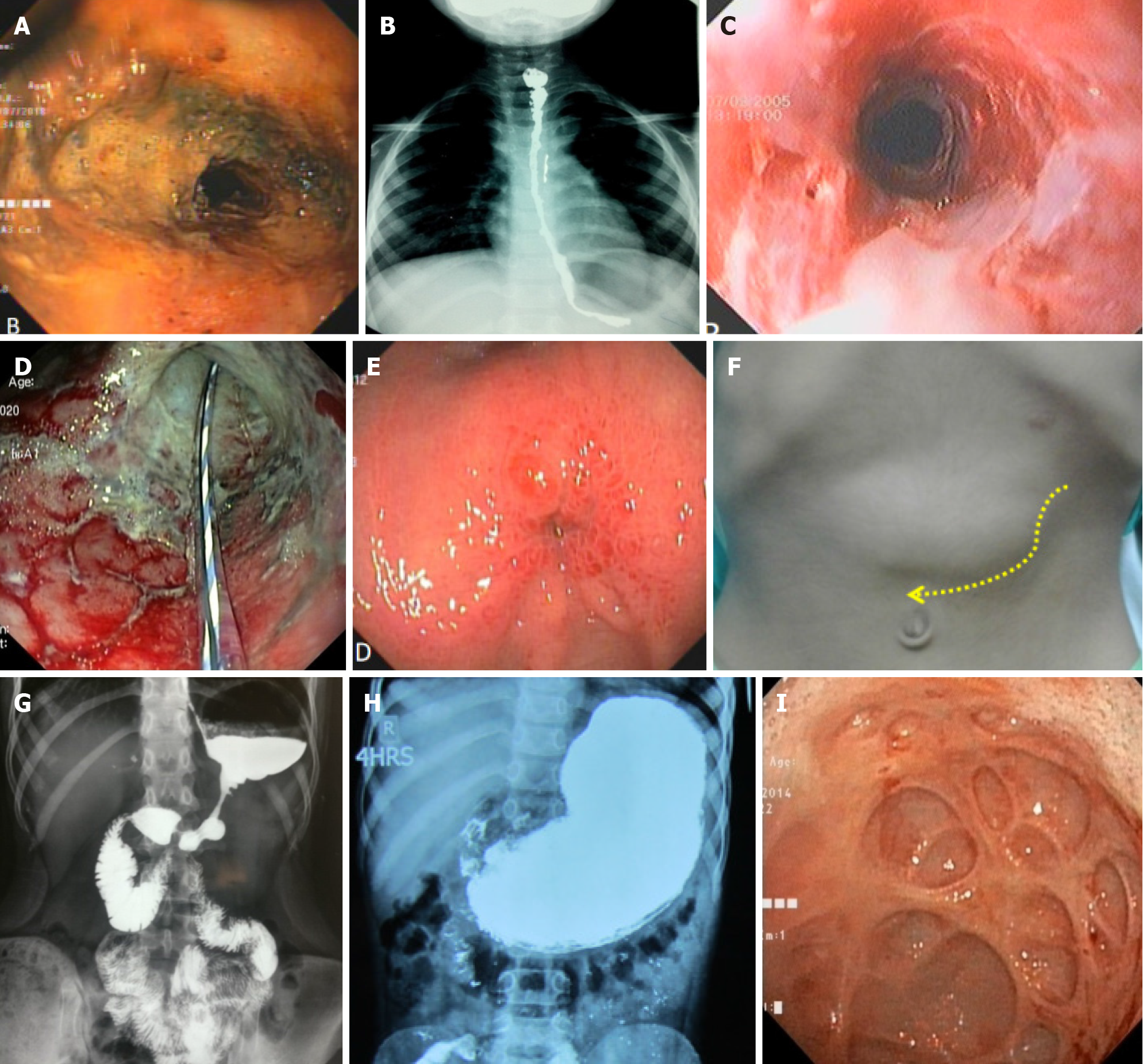
Figure 2 Clinical, endoscopic and radiological images of corrosive injury in children.
A: Endoscopic view of corrosive injury of esophagus (areas of necrosis); B: Barium swallow study showing long esophageal stricture; C: Endoscopic view of esophagus after initial healing; D: Endoscopic view of post-acid ingestion antropyloric injury with transpyloric tube in situ; E: Endoscopic view of pyloric stricture; F: Dilated stomach in a patient with pyloric stricture; G: Barium meal follow-through study showing corrosive stricture involving body and prepyloric region (Hour-glass appearnce); H: Barium meal follow through study showing post-corrosive pyloric stricture; I: Endoscopic view of diverticulae in stomach in pyloric stricture.
- Citation: Sarma MS, Tripathi PR, Arora S. Corrosive upper gastrointestinal strictures in children: Difficulties and dilemmas. World J Clin Pediatr 2021; 10(6): 124-136
- URL: https://www.wjgnet.com/2219-2808/full/v10/i6/124.htm
- DOI: https://dx.doi.org/10.5409/wjcp.v10.i6.124


