Copyright
©The Author(s) 2019.
World J Stomatol. Dec 18, 2019; 7(3): 28-38
Published online Dec 18, 2019. doi: 10.5321/wjs.v7.i3.28
Published online Dec 18, 2019. doi: 10.5321/wjs.v7.i3.28
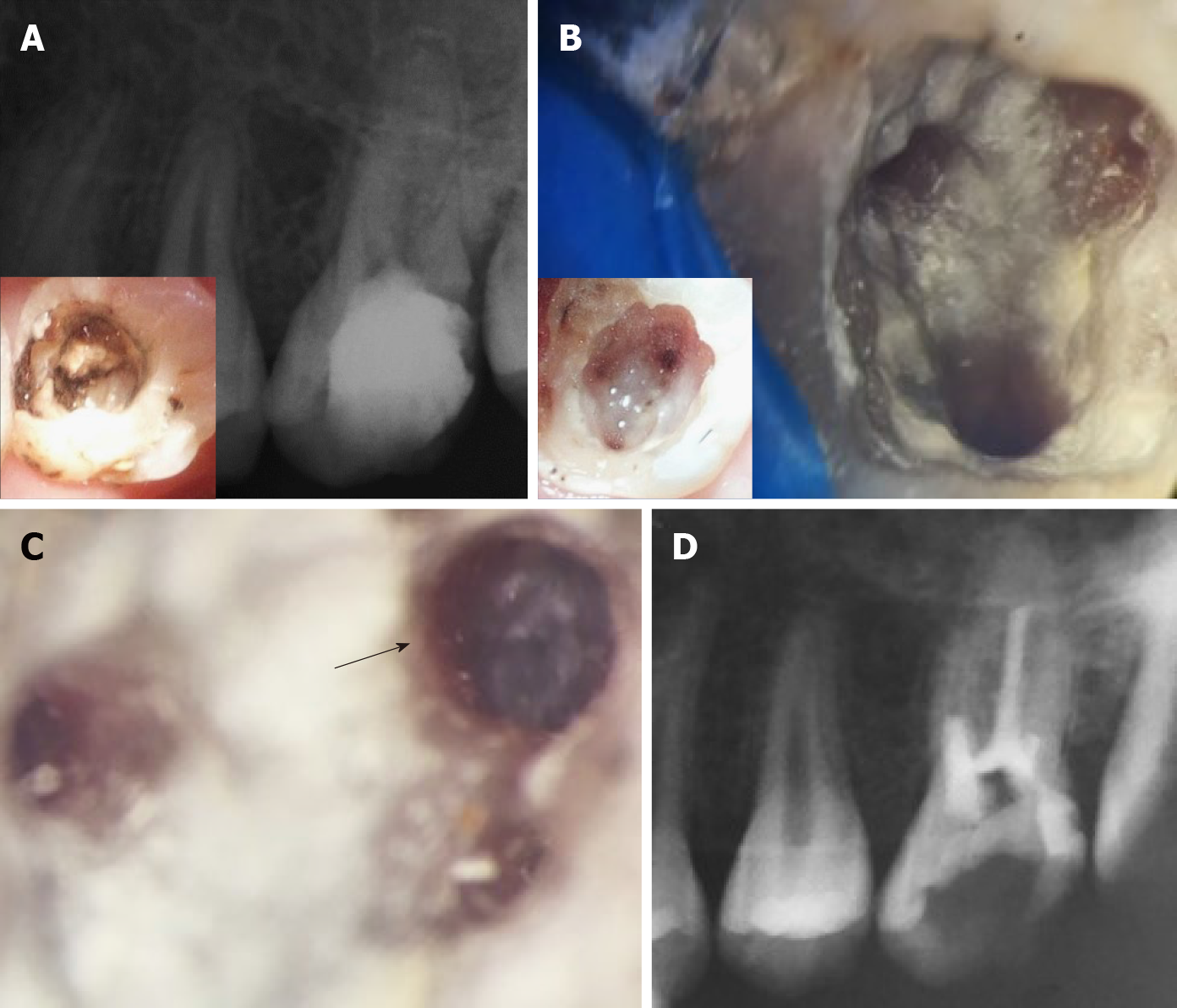
Figure 5 Four-year treatment outcome and re-treatment procedures for case #2 (tooth #14).
Figure shows the condition of the tooth after 4 years. A: The 4 years periapical radiograph showing recurrent decay on the distal surface of the tooth underneath the composite restoration. Insert in A shows the extent of the decay and the pulp chamber prior to complete removal of the MTA coronal plug. Note that the periapical tissues appear healthy; B: Shows the pulp chamber after removal of residual MTA material taken using a Kaaps dental operating microscope (x 10, Original magnification). Insert in B shows location of orifices. Minimal traces of fragmented tissues were retrieved from the pulp chamber; C: Higher magnification of the first mesiobuccal canal (x 25, original magnification) shows diffuse calcification and an obliterated canal beyond this level; D: Immediate post-operative radiograph showing root canal filling with gutta percha till the level of canal obliteration for the 4 canals. The maximum length reached was 16 mm for the palatal canal, 14 mm for the first mesiobuccal canal and 3 mm for the distobuccal canal while only the canal orifice was negotiated for the second mesiobuccal canal.
- Citation: Eltawila AM, El Backly R. Autologous platelet-rich-fibrin-induced revascularization sequelae: Two case reports. World J Stomatol 2019; 7(3): 28-38
- URL: https://www.wjgnet.com/2218-6263/full/v7/i3/28.htm
- DOI: https://dx.doi.org/10.5321/wjs.v7.i3.28


