Copyright
©The Author(s) 2019.
World J Stomatol. Dec 18, 2019; 7(3): 28-38
Published online Dec 18, 2019. doi: 10.5321/wjs.v7.i3.28
Published online Dec 18, 2019. doi: 10.5321/wjs.v7.i3.28
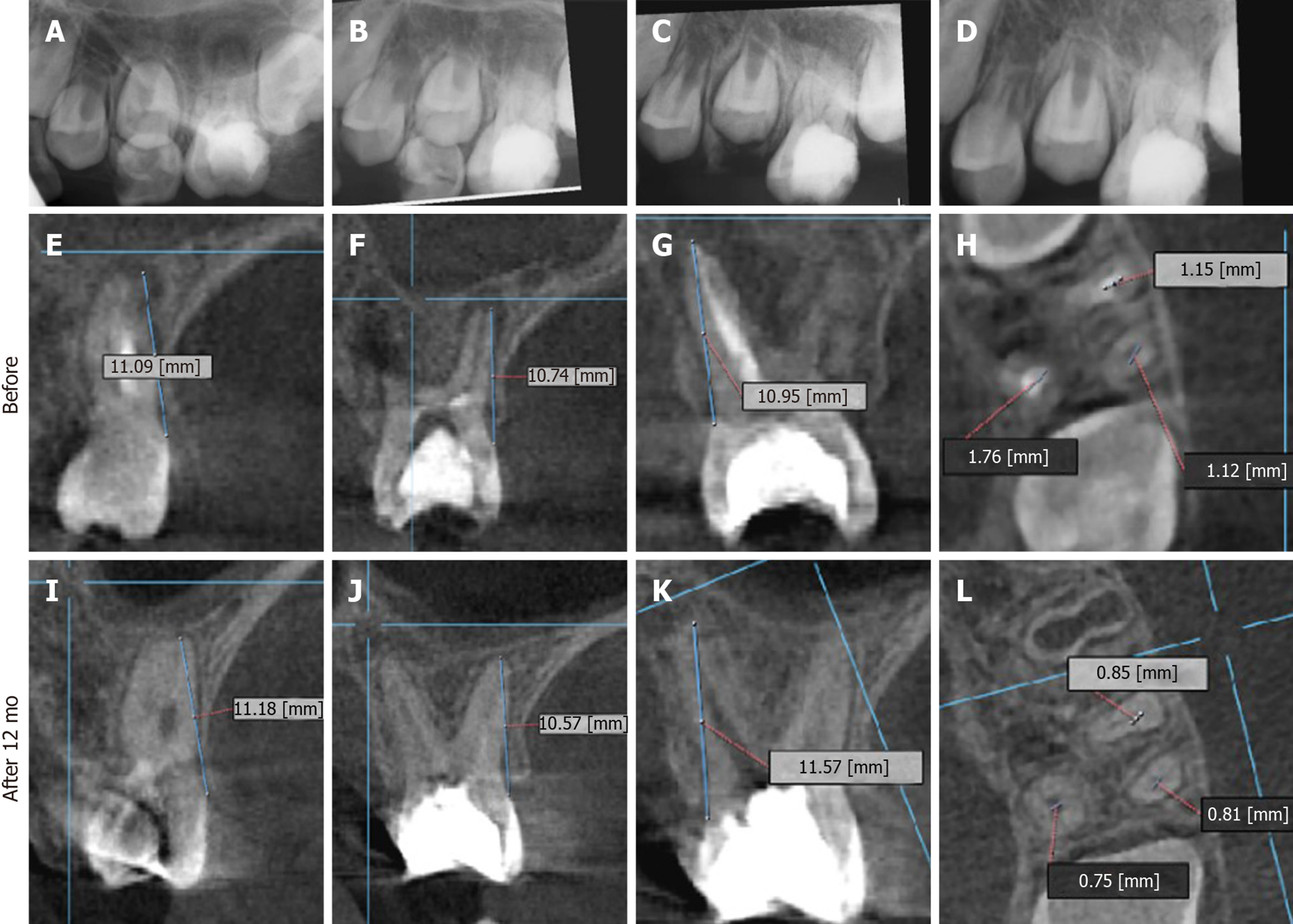
Figure 4 Twelve months periapical radiographs and cone beam computed tomography images of case #2 (tooth #14).
A-D: Digital periapical radiographs showing in an immediate post-operative radiograph; B: After 3 mo; C: After 6 mo and; D: After 12 mo; E-H: Cone beam computed tomography images (CBCT) images before the revascularization procedure (but after placement of intracanal medication); I-L: Follow-up after 12 mo; E, I: Mesial root; F, J: Distal root; G, K: Palatal root; H, L: Coronal section at apical third level showing the diameters of each root canal. All roots showed apical closure with complete periapical bone healing. Again, progress of periapical healing is evident along with root thickening and closure. The tooth remained asymptomatic throughout the follow-up period. All digital periapical radiographs were aligned using the Turboreg plugin- image J software (http://bigwww.epfl.ch/thevenaz/turboreg/). Details on the CBCT parameters and method are available in the supplementary material.
- Citation: Eltawila AM, El Backly R. Autologous platelet-rich-fibrin-induced revascularization sequelae: Two case reports. World J Stomatol 2019; 7(3): 28-38
- URL: https://www.wjgnet.com/2218-6263/full/v7/i3/28.htm
- DOI: https://dx.doi.org/10.5321/wjs.v7.i3.28


