Copyright
©The Author(s) 2019.
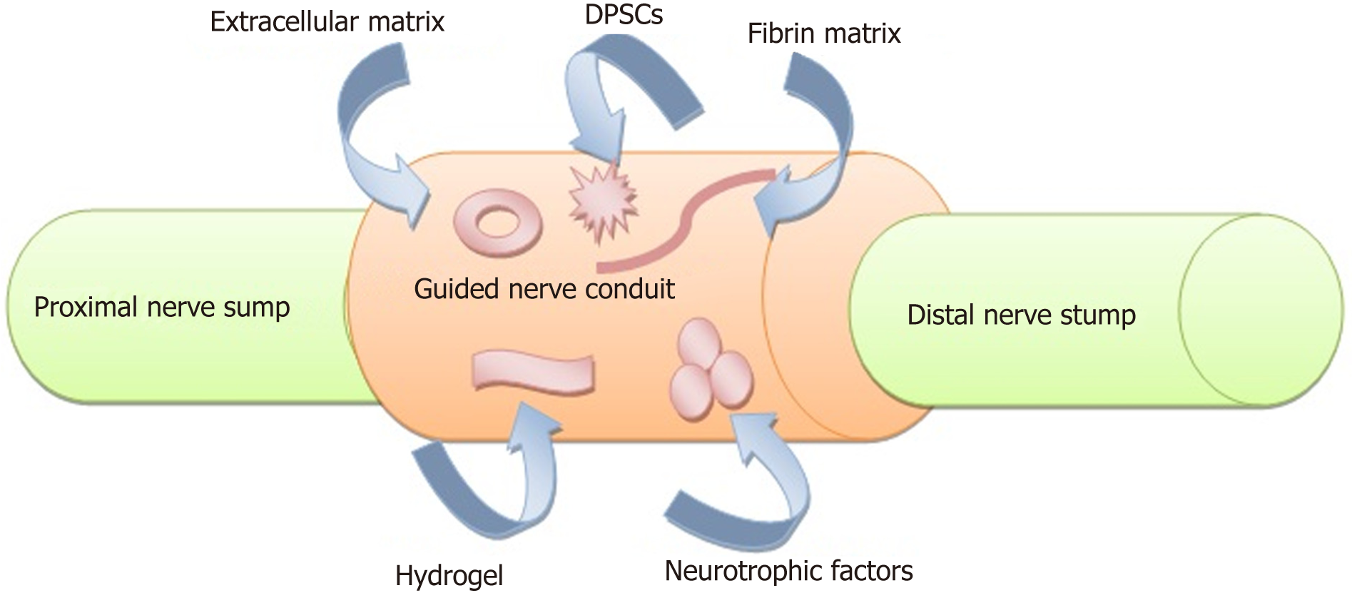
Figure 2 Schematic diagram showing a strategy to promote regeneration of bisection peripheral nerve injury using guided nerve conduit with incorporation of dental pulp stem cells and neurotrophic factors.
The proximal and distal nerve stumps (light green color) are sutured into the two ends of artificial nerve conduit (peach color). The conduit mimics the structures and components of autologous nerves and bridging the nerve gap to support the growth and regeneration of neural cells. The microenvironment of the conduit should contain nutrients, cytokines and growth factors (extracellular matrix, hydrogel and neurotrophic factors) as well as cellular elements (dental pulp stem cells).
- Citation: Sultan N, Amin LE, Zaher AR, Scheven BA, Grawish ME. Dental pulp stem cells: Novel cell-based and cell-free therapy for peripheral nerve repair. World J Stomatol 2019; 7(1): 1-19
- URL: https://www.wjgnet.com/2218-6263/full/v7/i1/1.htm
- DOI: https://dx.doi.org/10.5321/wjs.v7.i1.1


