Copyright
©2013 Baishideng Publishing Group Co.
World J Stomatol. Nov 20, 2013; 2(4): 103-107
Published online Nov 20, 2013. doi: 10.5321/wjs.v2.i4.103
Published online Nov 20, 2013. doi: 10.5321/wjs.v2.i4.103
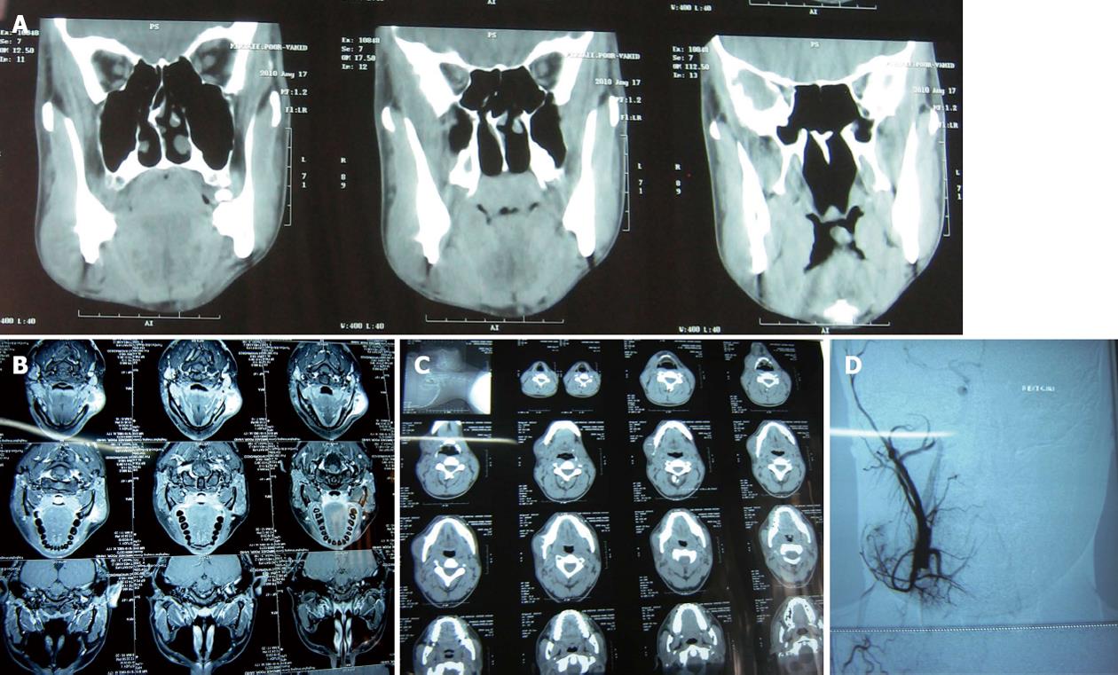
Figure 2 Computed tomography, magnetic resonance imaging and angiography.
A: Coronal-axial computed tomography scan showed a lesion appeared to have a non-uniform intra lesional; B: Magnetic resonance imaging (MRI), T1 post contrast view demonstrated a well defined lesion with high signal intensity in the superficial part of right mandibular body and angle; C: MRI, T2 post contrast view demonstrated a well defined lesion with high signal intensity in the superficial part of right mandibular body and angle; D: Angiography of right carotid artery showed a lesion with only mild to moderate vascularity and ruled out arteriovenous malformation and hemangioma.
- Citation: Jahanbani J, Sadri D, Hassani A, Kavandi F. Soft tissue aneurysmal bone cyst of the mandible: Report of a case. World J Stomatol 2013; 2(4): 103-107
- URL: https://www.wjgnet.com/2218-6263/full/v2/i4/103.htm
- DOI: https://dx.doi.org/10.5321/wjs.v2.i4.103


