Copyright
©2013 Baishideng Publishing Group Co.
World J Respirol. Jul 28, 2013; 3(2): 38-43
Published online Jul 28, 2013. doi: 10.5320/wjr.v3.i2.38
Published online Jul 28, 2013. doi: 10.5320/wjr.v3.i2.38
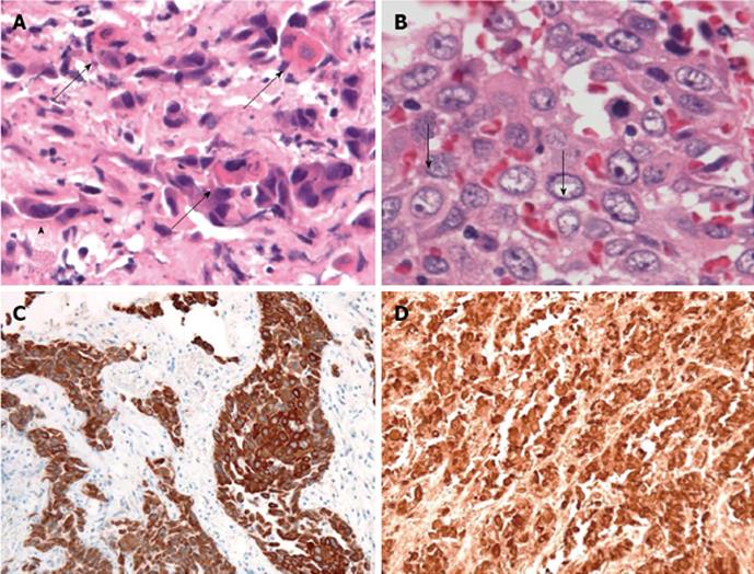
Figure 2 Histology of squamous cell carcinoma in the femur tumor.
A: Single cell keratosis (arrows); tadpole cells (arrowhead; HE staining, × 40); B: Intercellular bridges (arrows; HE staining, × 40); C: Cytokeratin 5/6 immunostaining (× 4) (monoclonal mouse anti-human cytokeratin 5/6 antibody from clone D5/16 B4; DakoCytomation, Copenhagen, Denmark); D: p63 immunostaining (× 4) (Monoclonal mouse anti-human p63 antibody from Clone 4A4; Nichirei Biosciences Inc., Tokyo, Japan).
-
Citation: Kanaji N, Bandoh S, Hayashi T, Haba R, Watanabe N, Ishii T, Kunitomo A, Takahama T, Tadokoro A, Imataki O, Dobashi H, Matsunaga T.
EGFR mutation identifies distant squamous cell carcinoma as metastasis from lung adenocarcinoma. World J Respirol 2013; 3(2): 38-43 - URL: https://www.wjgnet.com/2218-6255/full/v3/i2/38.htm
- DOI: https://dx.doi.org/10.5320/wjr.v3.i2.38


