Copyright
©The Author(s) 2021.
World J Respirol. Aug 8, 2021; 11(1): 12-17
Published online Aug 8, 2021. doi: 10.5320/wjr.v11.i1.12
Published online Aug 8, 2021. doi: 10.5320/wjr.v11.i1.12
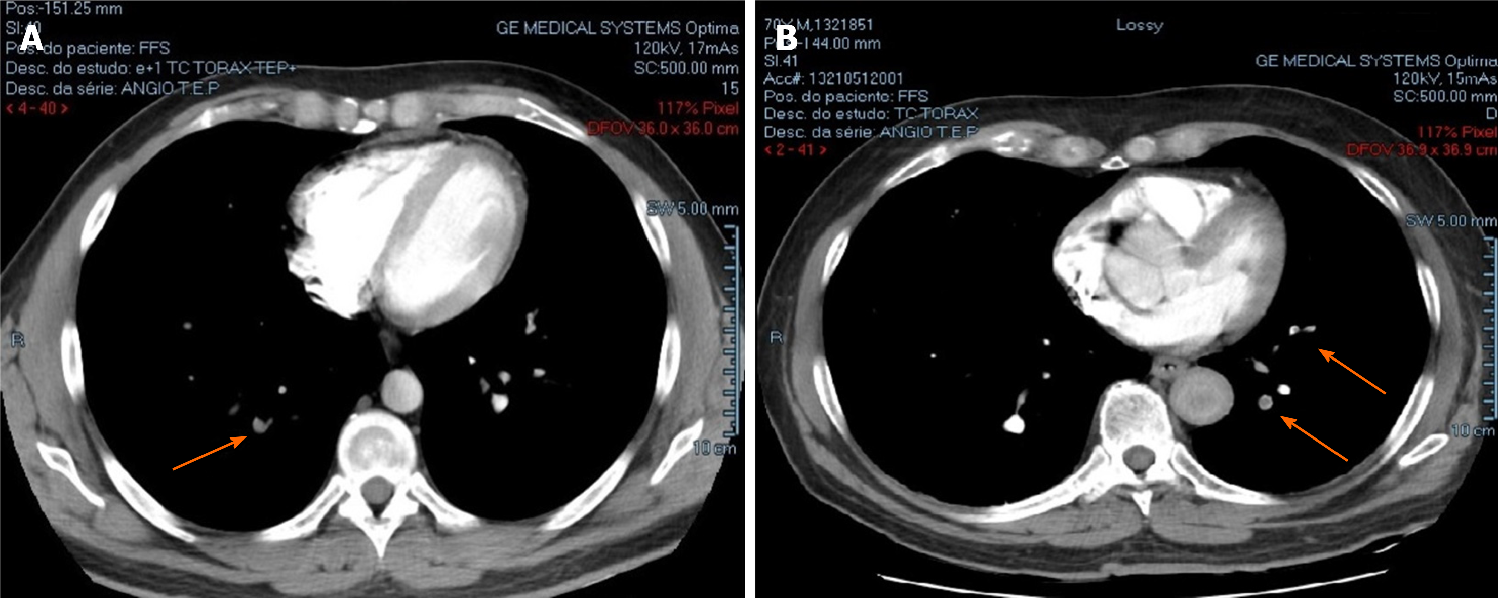
Figure 1 Pulmonary computed tomography angiography.
A: Father’s pulmonary computed tomography angiography transversal slice of lungs showed filling defects in subsegmental arterial branches characterizing left pulmonary embolism (arrows); B: Son’s pulmonary computed tomography angiography transversal slice of lungs showed right pulmonary embolism (arrow).
- Citation: Hannun P, Hannun W, Yoo HH, Resende L. Like father, like son: Pulmonary thromboembolism due to inflammatory or hereditary condition? Two case reports. World J Respirol 2021; 11(1): 12-17
- URL: https://www.wjgnet.com/2218-6255/full/v11/i1/12.htm
- DOI: https://dx.doi.org/10.5320/wjr.v11.i1.12


