Copyright
©The Author(s) 2021.
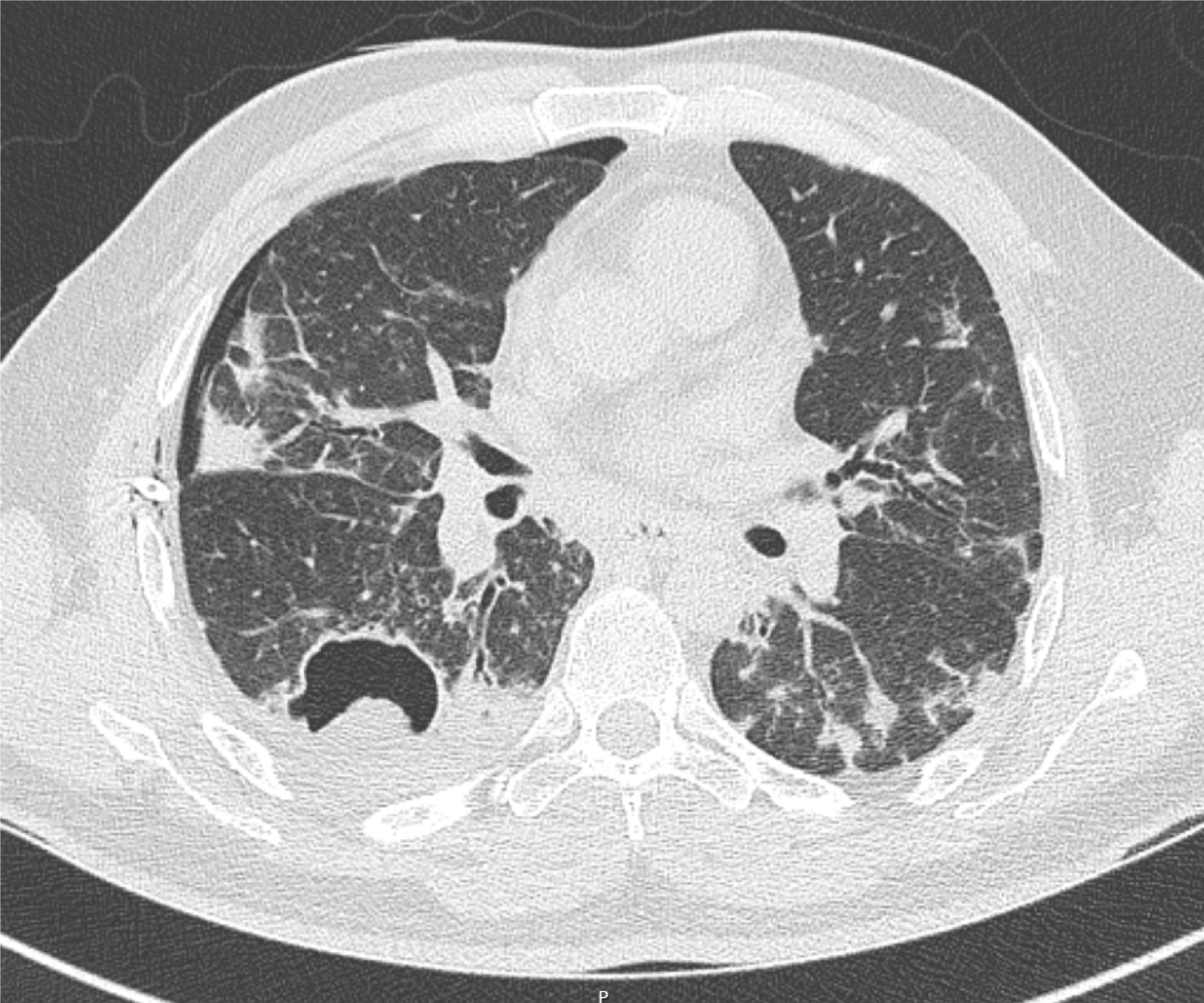
Figure 2 Computed tomography thorax image.
The thorax image depicting thin wall cavitary lesion with a fungal ball in the superior segment of the right lower lobe, right pneumothorax, and diffuse ground glass opacities and densities in bilateral lungs.
- Citation: Mathew J, Cherukuri SV, Dihowm F. SARS-CoV-2 with concurrent coccidioidomycosis complicated by refractory pneumothorax in a Hispanic male: A case report and literature review. World J Respirol 2021; 11(1): 1-11
- URL: https://www.wjgnet.com/2218-6255/full/v11/i1/1.htm
- DOI: https://dx.doi.org/10.5320/wjr.v11.i1.1


