Copyright
©The Author(s) 2019.
World J Otorhinolaryngol. Dec 20, 2019; 8(2): 12-18
Published online Dec 20, 2019. doi: 10.5319/wjo.v8.i2.12
Published online Dec 20, 2019. doi: 10.5319/wjo.v8.i2.12
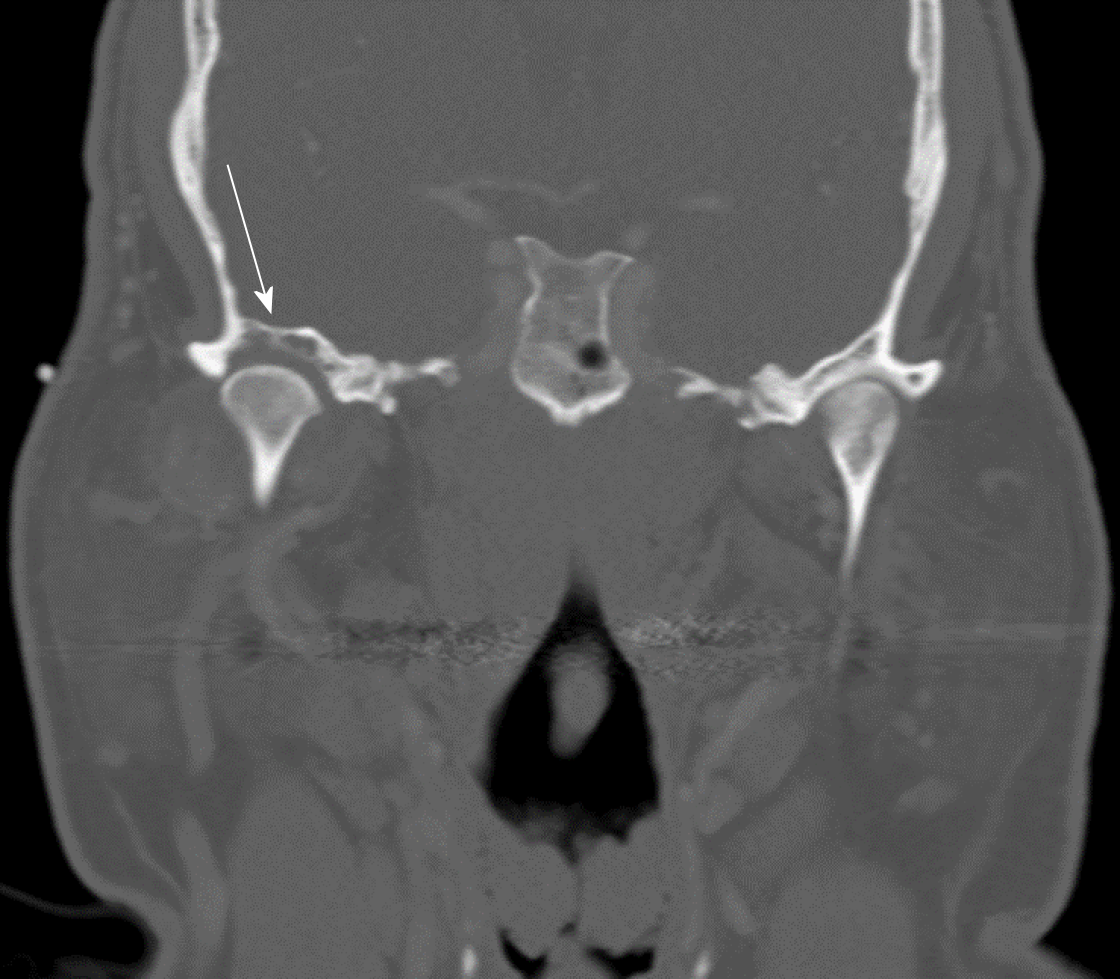
Figure 1 Coronal cut of computed tomography image (bone window) highlighting the erosion of the right glenoid fossa (arrow).
A soft tissue mass can faintly be observed on either side of the mandibular condyle, which is better delineated in a soft tissue window (not pictured).
- Citation: Romero N, Mulcahy CF, Barak S, Shand MF, Badger CD, Joshi AS. Synovial osteochondromatosis of the temporomandibular joint: A case report. World J Otorhinolaryngol 2019; 8(2): 12-18
- URL: https://www.wjgnet.com/2218-6247/full/v8/i2/12.htm
- DOI: https://dx.doi.org/10.5319/wjo.v8.i2.12


