Published online Aug 28, 2016. doi: 10.5319/wjo.v6.i3.50
Peer-review started: March 1, 2016
First decision: April 15, 2016
Revised: May 12, 2016
Accepted: June 1, 2016
Article in press: June 3, 2016
Published online: August 28, 2016
Processing time: 176 Days and 1.8 Hours
We describe a modified aural meatoplasty technique. The technique has been mainly used for mastoid surgery but it may also be used to address other causes of meatal stenosis. It involves removing most of the cartilage in the conchal bowl and soft tissue in the external auditory meatus. Cartilage from the helical root may also be sacrificed as part of this procedure. Our technique produces an excellent cosmetic result and an adequate meatoplasty which is easy to monitor in the outpatient setting.
Core tip: A successful meatoplasty addresses the bony, soft tissue and cartilaginous portions of the external auditory meatus. At least one, if not all these factors, can contribute to external auditory canal stenosis. Cartilage from the helical root can be resected in order to create an adequate meatoplasty.
- Citation: Kanzara T, Virk JS, Owa AO. Meatoplasty: A novel technique and minireview. World J Otorhinolaryngol 2016; 6(3): 50-53
- URL: https://www.wjgnet.com/2218-6247/full/v6/i3/50.htm
- DOI: https://dx.doi.org/10.5319/wjo.v6.i3.50
Creating an adequate meatoplasty is a critical step in allowing access and ventilation of the ear canal. Meatoplasty is often performed in conjunction with mastoid surgery. To create a successful meatoplasty, the surgeon considers the 3 main factors that contribute to meatal stenosis: (1) excess skin and soft tissue; (2) aberrations in the anatomy of the cartilage; or (3) the bony tympanic ring[1]. A range of techniques and variations have been reported in the literature since the original descriptions in 1893 by Stacke and Shwartze[2].
We present a technique developed at our centre by the senior author to ensure suitable meatoplasty size whilst maintaining cosmesis, principally in conjunction with mastoid surgery.
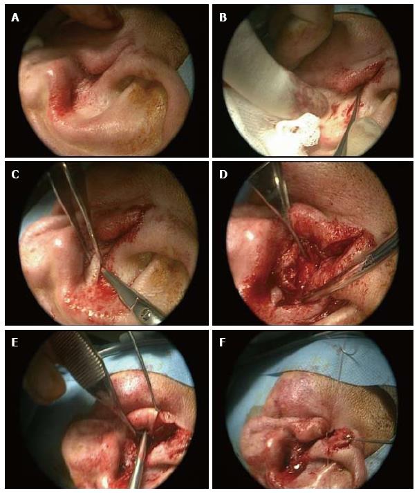
The steps include performing an endaural incision to bone (Figure 1A), followed by a skin incision over the superior aspect of the conchal bowl/cartilage (Figure 1B). A large portion of conchal cartilage is then removed, which can be used as part of the reconstruction in tympanomastoid surgery (Figure 1C). To facilitate the meatoplasty redundant mucosa and soft tissue is excised medial to the skin incision. An important factor is removal of prominent cartilage, for example at the root of the helix. The resultant skin flap is rotated into the cavity such that the meatoplasty is widened. Following this, absorbable sutures are used to ensure the tragus remains anterosuperior, by suturing the anterior edge of the endaural incision (Figure 1D). The free edge of conchal skin is opposed with absorbable suture (Figure 1E and F). An adequately sized meatoplasty should not require splinting by packing such as with bismuth iodoform paraffin paste (BIPP).
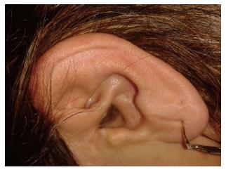
This technique for meatoplasty over the preceding two years has resulted in very good functional and cosmetic outcomes in all 54 patients (Figure 2). We have managed to achieve a dry, auto cleaning ear with very good cosmetic results in all cases. We have not had any cases of stenosis or troublesome granulation. Therefore, we propose our technique as a useful adaptation in performing meatoplasty.
There is an abundance of meatoplasty techniques in the literature reportedly producing very good results in the respective author’s hands. These techniques rely on creating a meatoplasty using a fibrocartilaginous posterior meatal flap in a post-auricular approach mastoidectomy, or partially resecting conchal bowl cartilage for the endaural approach[3]. Traditional thinking with regards to meatoplasty surgery has tended to focus on creating a large meatoplasty thought to support optimal ventilation, reduce both bacterial and debris accumulation[4-7]. However, the “big is better” approach is often associated with a few complications which include poor cosmesis and poor hearing aid fitting amongst other things[8,9].
Two broad approaches have been reported with regards to widening the skin of the entrance of the external auditory meatus. The first approach, which in essence forms the basis of the Portman technique, uses radial incisions to create flaps arising from the outer ring of the external auditory meatus[10]. Following the excision of underlying soft tissue, the flaps are then elevated onto the bony canal. The second approach, originally described by Korner and Siebenmann but reported by others, involves dividing the ring and rotating a skin flap to cover the defect[1,11].
More recent techniques, similarly ours, have tended to favour the second approach involving rotational flaps. Osborne and Martin[12] described rotating a superiorly placed pre-tragal flap into the endaural incision following removal of some conchal bowl cartilage. Hovis et al[13] described a “one cut meatoplasty” involving making a single horizontal incision from the posterosuperior meatus to the antihelix to create an inferiorly based triangular conchal flap. Martin-Hirsch and Smelt described a flap starting medial to the excess conchal mucosa and extending superiorly[14]. Follow-up results of patients managed with this technique over a 9-year period have shown favourable results especially for the otitis externa group[15]. Banerjee et al[16] reported using an inferiorly based transposition flap which potentially has poor cosmesis as its major demerit owing to the inferoposterior widening inherent in the technique[1,14].
Other techniques which do not make use of skin flaps have been reported. Patil et al[1] describe a technique which involves the removal of a small triangle of skin on the entrance of the external auditory canal in addition to conchal cartilage resection. Goodyear et al[17] described a helical advancement technique which involves resecting a cuff of skin anterior to the helix followed by releasing the helix anteriorly to cover the defect. The authors point out that whilst this approach produces inferior cosmesis it has the advantage of producing an auto-cleaning cavity which is easy to manage in the outpatient setting[17].
In our experience of using this technique, we have not experienced any of the common complications associated with meatoplasty such as perichondritis, discharge or restenosis. Further, the patients operated on have not complained about poor cosmesis on follow-up.
We believe this previously undescribed methodology will provide an additional tool in the armamentarium of the otolaryngologist. Our modified approach demonstrated in this paper is reproducible and technically straightforward. It provides an adequate meatoplasty which promotes an auto cleaning ear that is easily accessible in clinic whilst producing an excellent cosmetic result.
Manuscript source: Invited manuscript
Specialty type: Otorhinolaryngology
Country of origin: United Kingdom
Peer-review report classification
Grade A (Excellent): 0
Grade B (Very good): B
Grade C (Good): C
Grade D (Fair): 0
Grade E (Poor): 0
P- Reviewer: Pan F, Vynios D S- Editor: Ji FF L- Editor: A E- Editor: Lu YJ
| 1. | Patil S, Ahmed J, Patel N. Endaural meatoplasty: the Whipps Cross technique. J Laryngol Otol. 2011;125:78-81. [PubMed] [DOI] [Cited in This Article: ] [Cited by in Crossref: 4] [Cited by in F6Publishing: 5] [Article Influence: 0.4] [Reference Citation Analysis (1)] |
| 2. | Roulleau P. [Development of the surgery of chronic otitis]. Rev Laryngol Otol Rhinol (Bord). 1993;114:87-91. [PubMed] [Cited in This Article: ] |
| 3. | Phillips WC. The radical mastoid operation. Diseases of the Ear, Nose and Throat Medical and Surgical. Philadelphia: FA Davis 1929; 311-343. [Cited in This Article: ] |
| 4. | Jackson CG, Glasscock ME, Nissen AJ, Schwaber MK, Bojrab DI. Open mastoid procedures: contemporary indications and surgical technique. Laryngoscope. 1985;95:1037-1043. [PubMed] [DOI] [Cited in This Article: ] [Cited by in Crossref: 32] [Cited by in F6Publishing: 34] [Article Influence: 0.9] [Reference Citation Analysis (0)] |
| 5. | Nadol JB. Revision mastoidectomy. Otolaryngol Clin North Am. 2006;39:723-740, vi-vii. [PubMed] [DOI] [Cited in This Article: ] [Cited by in Crossref: 25] [Cited by in F6Publishing: 27] [Article Influence: 1.5] [Reference Citation Analysis (0)] |
| 6. | Sheehy JL. Cholesteatoma surgery: canal wall down procedures. Ann Otol Rhinol Laryngol. 1988;97:30-35. [PubMed] [DOI] [Cited in This Article: ] [Cited by in Crossref: 43] [Cited by in F6Publishing: 45] [Article Influence: 1.3] [Reference Citation Analysis (0)] |
| 7. | Pillsbury HC, Carrasco VN. Revision mastoidectomy. Arch Otolaryngol Head Neck Surg. 1990;116:1019-1022. [PubMed] [DOI] [Cited in This Article: ] [Cited by in Crossref: 22] [Cited by in F6Publishing: 23] [Article Influence: 0.7] [Reference Citation Analysis (0)] |
| 8. | Osborne JE, Terry RM, Gandhi AG. Large meatoplasty technique for mastoid cavities. Clin Otolaryngol Allied Sci. 1985;10:357-360. [PubMed] [DOI] [Cited in This Article: ] [Cited by in Crossref: 19] [Cited by in F6Publishing: 20] [Article Influence: 0.5] [Reference Citation Analysis (0)] |
| 9. | Gluth MB, Friedman AB, Atcherson SR, Dornhoffer JL. Hearing aid tolerance after revision and obliteration of canal wall down mastoidectomy cavities. Otol Neurotol. 2013;34:711-714. [PubMed] [DOI] [Cited in This Article: ] [Cited by in Crossref: 14] [Cited by in F6Publishing: 15] [Article Influence: 1.4] [Reference Citation Analysis (0)] |
| 10. | Portmann M. “How I do it”--otology and neurotology. A specific issue and its solution. Meatoplasty and conchoplasty in cases of open technique. Laryngoscope. 1983;93:520-522. [PubMed] [Cited in This Article: ] |
| 11. | Wormald PJ, van Hasselt CA. A technique of mastoidectomy and meatoplasty that minimizes factors associated with a discharging mastoid cavity. Laryngoscope. 1999;109:478-482. [PubMed] [DOI] [Cited in This Article: ] [Cited by in Crossref: 11] [Cited by in F6Publishing: 13] [Article Influence: 0.5] [Reference Citation Analysis (0)] |
| 12. | Osborne JE, Martin FW. Endaural meatoplasty for mastoid cavities. Clin Otolaryngol Allied Sci. 1990;15:453-455. [PubMed] [DOI] [Cited in This Article: ] [Cited by in Crossref: 8] [Cited by in F6Publishing: 9] [Article Influence: 0.3] [Reference Citation Analysis (0)] |
| 13. | Hovis KL, Carlson ML, Sweeney AD, Haynes DS. The one-cut meatoplasty: novel surgical technique and outcomes. Am J Otolaryngol. 2015;36:130-135. [PubMed] [DOI] [Cited in This Article: ] [Cited by in Crossref: 5] [Cited by in F6Publishing: 6] [Article Influence: 0.7] [Reference Citation Analysis (0)] |
| 14. | Martin-Hirsch DP, Smelt GJ. Conchal flap meatoplasty. J Laryngol Otol. 1993;107:1029-1031. [PubMed] [DOI] [Cited in This Article: ] [Cited by in Crossref: 6] [Cited by in F6Publishing: 7] [Article Influence: 0.2] [Reference Citation Analysis (0)] |
| 15. | Kumar PJ, Smelt GJ. A long term follow up of conchal flap meatoplasty in chronic otitis externa. J Laryngol Otol. 2007;121:1-4. [PubMed] [Cited in This Article: ] |
| 16. | Banerjee AR, Moir AA, Jervis P, Narula AA. A canalplasty technique for the surgical treatment of chronic otitis externa. Clin Otolaryngol Allied Sci. 1995;20:150-152. [PubMed] [DOI] [Cited in This Article: ] |
| 17. | Goodyear PW, Reddy CE, Lesser TH. Helix advancement meatoplasty. J Laryngol Otol. 2012;126:612-614. [PubMed] [DOI] [Cited in This Article: ] [Cited by in Crossref: 3] [Cited by in F6Publishing: 4] [Article Influence: 0.3] [Reference Citation Analysis (0)] |


