Copyright
©The Author(s) 2016.
World J Otorhinolaryngol. Feb 28, 2016; 6(1): 13-18
Published online Feb 28, 2016. doi: 10.5319/wjo.v6.i1.13
Published online Feb 28, 2016. doi: 10.5319/wjo.v6.i1.13
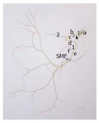
Figure 1 Line drawing of the facial nerve.
The nerve takes a very tortuous course through the temporal bone within the Fallopian canal before exiting the skull base at the SMF. Any portion, intra or extratemporal, can be affected by a facial nerve schwannoma. a: Nerve as it exits the brainstem and crosses the cerebellopontine angle; b: Labyrinthine segment; c: Geniculate ganglion; d: Tympanic segment; e: Mastoid segment; f: Chorda tympani nerve; arrow: Intracanilicular segment. 1Nerve to stapedius; 2Greater superficial petrosal nerve. (Joshua M Klein, MSM I artist). SMF: Stylomastoid foramen.
- Citation: Makadia L, Mowry SE. Management of intratemporal facial nerve schwannomas: The evolution of treatment paradigms from 2000-2015. World J Otorhinolaryngol 2016; 6(1): 13-18
- URL: https://www.wjgnet.com/2218-6247/full/v6/i1/13.htm
- DOI: https://dx.doi.org/10.5319/wjo.v6.i1.13


