Copyright
©2014 Baishideng Publishing Group Inc.
World J Otorhinolaryngol. Nov 28, 2014; 4(4): 17-22
Published online Nov 28, 2014. doi: 10.5319/wjo.v4.i4.17
Published online Nov 28, 2014. doi: 10.5319/wjo.v4.i4.17
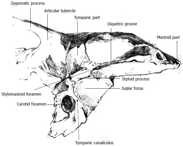
Figure 2 Illustration of the temporal bone demonstrating the relationship between the tympanic canaliculus, the carotid foramen and the jugular fossa.
- Citation: Kanzara T, Hall A, Virk JS, Leung B, Singh A. Clinical anatomy of the tympanic nerve: A review. World J Otorhinolaryngol 2014; 4(4): 17-22
- URL: https://www.wjgnet.com/2218-6247/full/v4/i4/17.htm
- DOI: https://dx.doi.org/10.5319/wjo.v4.i4.17


