Copyright
©2013 Baishideng Publishing Group Co.
World J Otorhinolaryngol. Nov 28, 2013; 3(4): 114-133
Published online Nov 28, 2013. doi: 10.5319/wjo.v3.i4.114
Published online Nov 28, 2013. doi: 10.5319/wjo.v3.i4.114
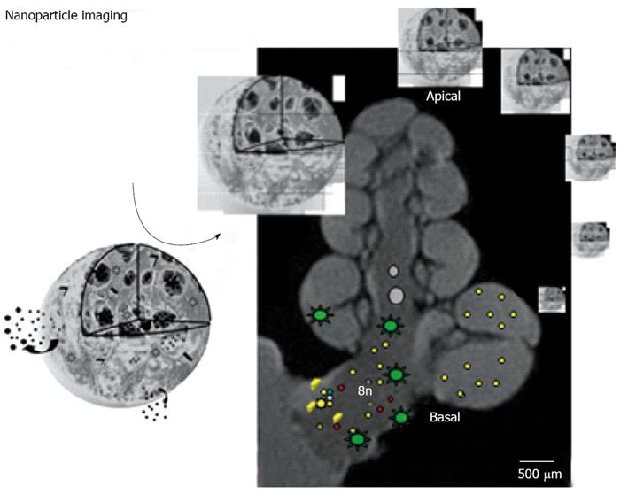
Figure 9 Schematic representation of imaging of nanoparticles in the inner ear of guinea pig with magnetic resonance imaging.
The cochlear nerve (8n) and cochlear basal turn (basal) are indicated. As nanoparticle transmission electron microscopy of multifunctional poly-lactic-co-glycolic acid -nanoparticle is shown. The star-like dots in the fluid spaces of the cochlea and the cochlear nerve demonstrate the distribution of gadolinium chelate used for visualization. The small dots indicate nanoparticles, their ingredients and dye for histological confirmation of targeting.
- Citation: Pyykkö I, Zou J, Zhang Y, Zhang W, Feng H, Kinnunen P. Nanoparticle based inner ear therapy. World J Otorhinolaryngol 2013; 3(4): 114-133
- URL: https://www.wjgnet.com/2218-6247/full/v3/i4/114.htm
- DOI: https://dx.doi.org/10.5319/wjo.v3.i4.114


