Copyright
©2013 Baishideng.
World J Otorhinolaryngol. Feb 28, 2013; 3(1): 1-15
Published online Feb 28, 2013. doi: 10.5319/wjo.v3.i1.1
Published online Feb 28, 2013. doi: 10.5319/wjo.v3.i1.1
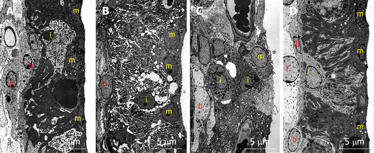
Figure 7 Transmission electron microscopy findings in the stria vascularis before and 1, 4, and 7 d after ischemic insult.
A: Marginal cells on the medial aspect of the stria vascularis showed extensive branching processes with intermediate cells. The basal cells, located on the lateral aspect of the stria vascularis, connected to the intermediate cells and type 1 fibrocytes of the spiral ligament by means of gap junctions; B: Vacuoles were seen in marginal cells. The intercellular space increased and intermediate cells seemed to have shrunk; C: Vacuoles persisted in marginal cells. The intermediate cells were still shrunken, although the intercellular space was smaller than that on day 1; D: The intercellular space was no longer enlarged, although a few small vacuoles were found in marginal cells. The extensive branching processes appeared to have a normal shape. m: Marginal cell; i: Intermediate cell; b: Basal cell; f: Type 1 fibrocyte.
- Citation: Gyo K. Experimental study of transient cochlear ischemia as a cause of sudden deafness. World J Otorhinolaryngol 2013; 3(1): 1-15
- URL: https://www.wjgnet.com/2218-6247/full/v3/i1/1.htm
- DOI: https://dx.doi.org/10.5319/wjo.v3.i1.1


