Copyright
©The Author(s) 2015.
World J Ophthalmol. May 12, 2015; 5(2): 86-98
Published online May 12, 2015. doi: 10.5318/wjo.v5.i2.86
Published online May 12, 2015. doi: 10.5318/wjo.v5.i2.86
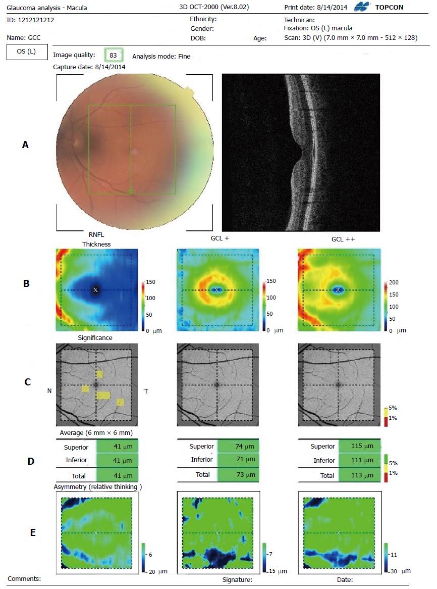
Figure 4 Three dimensions optical coherence tomography 2000.
A: Segmentation: 7 mm2 area centered on the fovea with a scan density of 512 vertical × 128 horizontal scans; B: Thickness map. Average regional thickness is calculated for RNFL, GCL+ (GCL + IPL), GCL++ (RNFL + GCL + IPL). Each cell is calculated and compared to the normative database of the device; C: Significance map. From left to right, 10 × 10 grid comparison maps covering 6 mm × 6 mm area of RNFL, GCL+ and GCL++ are shown. The comparison result is displayed with the color in the legend on the right. The background image is red free image; D: Average thickness. From left to right, three average thicknesses of RNFL, GCL+ and GCL++. The top is “Superior” which means average in the upper half area, the middle is “Inferior” which means average in the lower half area, and the bottom is “Total’ which means average in the total area. Each average thickness is compared to the normative data and displayed according to color; E: Asymmetry map. From left to right, subtraction thickness maps covering 6 mm × 6 mm area of RNFL, GCL+ and GCL++ are shown. The subtraction is performed between two points which symmetrically lie with respect to the center horizontal line. In the upper half, the value in each point is calculated such that thickness of the point is subtracted from the thickness of the corresponding line-symmetry point below and vice versa. Blue indicates that the thickness of the point is thinner than that of the corresponding point. RNFL: Retinal nerve fiber layer; GCL: Ganglion cell layer; IPL: Inner plexiform layer.
- Citation: Meshi A, Goldenberg D, Armarnik S, Segal O, Geffen N. Systematic review of macular ganglion cell complex analysis using spectral domain optical coherence tomography for glaucoma assessment. World J Ophthalmol 2015; 5(2): 86-98
- URL: https://www.wjgnet.com/2218-6239/full/v5/i2/86.htm
- DOI: https://dx.doi.org/10.5318/wjo.v5.i2.86


