Copyright
©2014 Baishideng Publishing Group Inc.
World J Ophthalmol. Nov 12, 2014; 4(4): 113-123
Published online Nov 12, 2014. doi: 10.5318/wjo.v4.i4.113
Published online Nov 12, 2014. doi: 10.5318/wjo.v4.i4.113
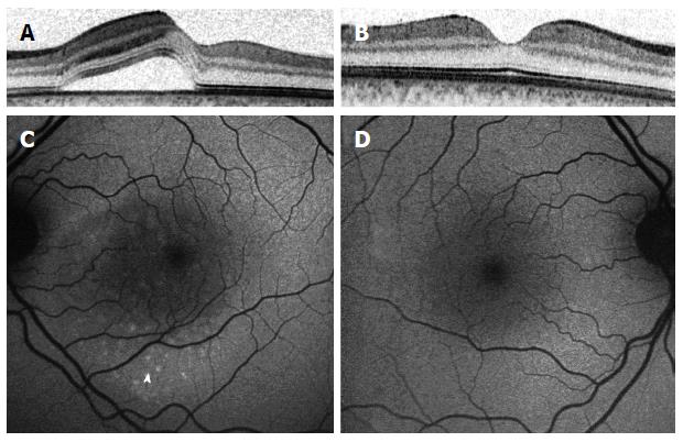
Figure 4 Imaging of a 38-year-old patient with central serous chorioretinopathy, one month following the beginning of his symptoms.
Comparison of left (A) and right (B) eyes imaged with spectral domain optical coherence tomography shows a serous retinal detachment in his left eye. Minimal changes are seen with fundus autofluorescence imaging (C), consisting of a granular pattern of increased autofluorescence in the area of retinal detachment (arrowhead), compared to images of the right eye (D).
- Citation: Schaap-Fogler M, Ehrlich R. What is new in central serous chorioretinopathy? World J Ophthalmol 2014; 4(4): 113-123
- URL: https://www.wjgnet.com/2218-6239/full/v4/i4/113.htm
- DOI: https://dx.doi.org/10.5318/wjo.v4.i4.113


