Copyright
©2014 Baishideng Publishing Group Inc.
World J Ophthalmol. Aug 12, 2014; 4(3): 56-62
Published online Aug 12, 2014. doi: 10.5318/wjo.v4.i3.56
Published online Aug 12, 2014. doi: 10.5318/wjo.v4.i3.56
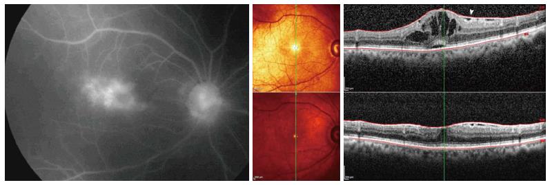
Figure 1 Uveitic macular edema.
Left: Late frame of fluorescein angiogram showing dye leakage to the macular area with cystoid pattern (cystoid macular edema) due to uveitis. Upper right: Spectral-domain optical coherence tomography orientation and B-scans of the same eye showing cystoid macular edema with small amount of subfoveal fluid and associated epiretinal membrane (white arrowhead). Lower right: Spectral-domain optical coherence tomography orientation and B-scans of the same eye 3 mo after intravitreal triamcinolone acetonide intravitreal injection demonstrates complete resolution of intraretinal and subfoveal fluid with persistence of epiretinal membrane. Visual acuity improved from 20/100 to 20/30.
- Citation: Shoughy SS, Kozak I. Updates in uveitic macular edema. World J Ophthalmol 2014; 4(3): 56-62
- URL: https://www.wjgnet.com/2218-6239/full/v4/i3/56.htm
- DOI: https://dx.doi.org/10.5318/wjo.v4.i3.56


