Copyright
©2014 Baishideng Publishing Group Inc.
World J Ophthalmol. Aug 12, 2014; 4(3): 35-46
Published online Aug 12, 2014. doi: 10.5318/wjo.v4.i3.35
Published online Aug 12, 2014. doi: 10.5318/wjo.v4.i3.35
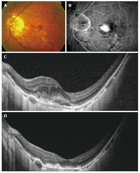
Figure 3 Resolution seen with anti-vascular endothelial growth factor treatment in myopic.
Choroidal neovascularization (CNV) color fundus photograph (A), fluorescein angiography (FA) (B) and optical coherence tomography (OCT) (C) showing a larger juxtafoveal myopic CNV with significant amount of intraretinal fluid. Note the lacquer cracks which are clearly visible on the FA. After a course of intravitreal bevacizumab, OCT demonstrated resolution of intraretinal fluid and consolidation of the CNV (D).
- Citation: Teo K, Cheung CMG. Choroidal neovascularization secondary to pathological myopia. World J Ophthalmol 2014; 4(3): 35-46
- URL: https://www.wjgnet.com/2218-6239/full/v4/i3/35.htm
- DOI: https://dx.doi.org/10.5318/wjo.v4.i3.35


