Copyright
©2014 Baishideng Publishing Group Inc.
World J Ophthalmol. Aug 12, 2014; 4(3): 35-46
Published online Aug 12, 2014. doi: 10.5318/wjo.v4.i3.35
Published online Aug 12, 2014. doi: 10.5318/wjo.v4.i3.35
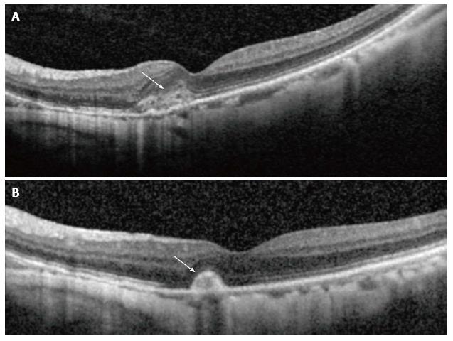
Figure 2 Optical coherence tomography showing corresponding changes in activity.
(A) Shows hyperreflective lesion corresponding to a small juxtafoveal myopic choroidal neovascularization (arrow) located above the retinal pigment epithelial cell layer with minimal exudation. After 3 mo (B), the lesion had scarred up, represented by a highly reflective lesion with sharp outline, and no intraretinal fluid is seen, which suggests an inactive lesion (arrow).
- Citation: Teo K, Cheung CMG. Choroidal neovascularization secondary to pathological myopia. World J Ophthalmol 2014; 4(3): 35-46
- URL: https://www.wjgnet.com/2218-6239/full/v4/i3/35.htm
- DOI: https://dx.doi.org/10.5318/wjo.v4.i3.35


