Copyright
©2014 Baishideng Publishing Group Inc.
World J Ophthalmol. Aug 12, 2014; 4(3): 35-46
Published online Aug 12, 2014. doi: 10.5318/wjo.v4.i3.35
Published online Aug 12, 2014. doi: 10.5318/wjo.v4.i3.35
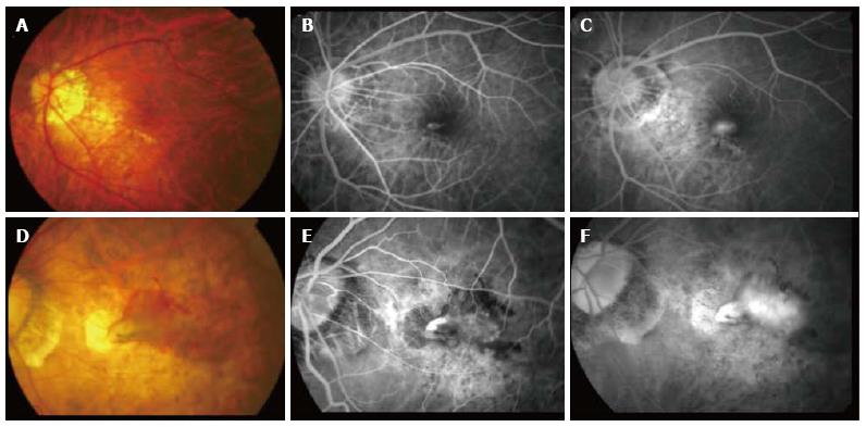
Figure 1 Clinical features of a typical choroidal neovascularization secondary to pathological myopia.
(A) Color fundus photography and corresponding fluorescein angiography (FA) (B and C) showing a younger patient with a small choroidal neovascularization lesion adjacent to the lacquer crack compared to (D) color fundus photograph and corresponding FA (E and F) showing an older patient with a much larger lesion with significant intraretinal fluid and extensive background atrophic changes.
- Citation: Teo K, Cheung CMG. Choroidal neovascularization secondary to pathological myopia. World J Ophthalmol 2014; 4(3): 35-46
- URL: https://www.wjgnet.com/2218-6239/full/v4/i3/35.htm
- DOI: https://dx.doi.org/10.5318/wjo.v4.i3.35


