Copyright
©2013 Baishideng Publishing Group Co.
World J Ophthalmol. Nov 12, 2013; 3(4): 38-40
Published online Nov 12, 2013. doi: 10.5318/wjo.v3.i4.38
Published online Nov 12, 2013. doi: 10.5318/wjo.v3.i4.38
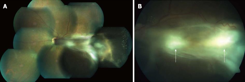
Figure 1 Fundus photo of patient’ right eye.
A: Montage colour fundus photo of patient showing extent of lesion with multifocal granulomata and temporal exudative retinal detachment; B: Fundus photo showing granulomata.
- Citation: Kuniyal L, Biswas J. Multifocal granulomata in presumed Toxocara canis infection in adult. World J Ophthalmol 2013; 3(4): 38-40
- URL: https://www.wjgnet.com/2218-6239/full/v3/i4/38.htm
- DOI: https://dx.doi.org/10.5318/wjo.v3.i4.38


