Copyright
©2011 Baishideng Publishing Group Co.
World J Ophthalmol. Dec 30, 2011; 1(1): 4-10
Published online Dec 30, 2011. doi: 10.5318/wjo.v1.i1.4
Published online Dec 30, 2011. doi: 10.5318/wjo.v1.i1.4
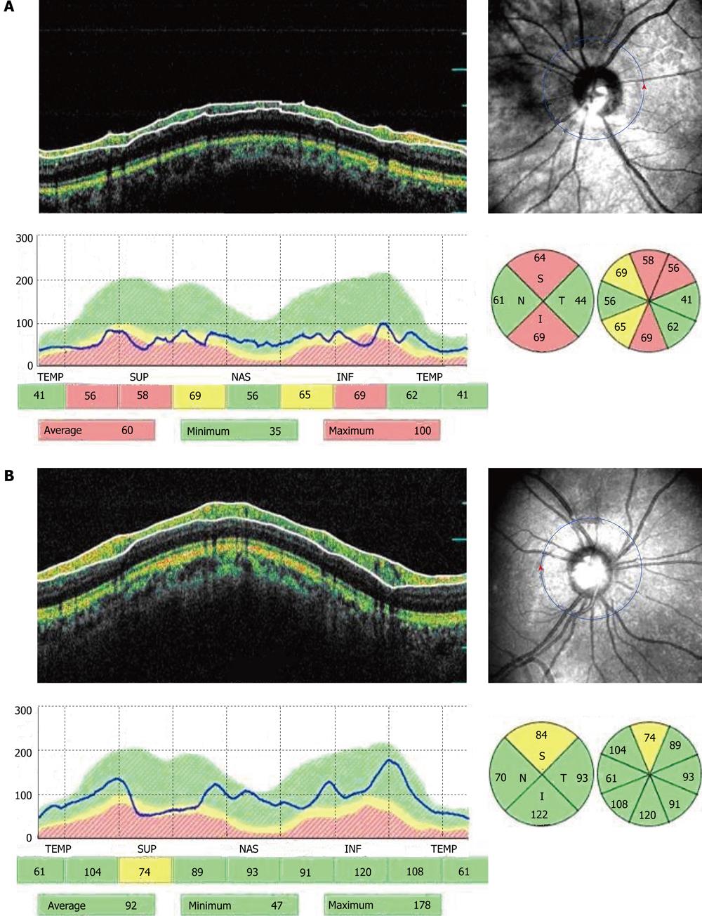
Figure 1 A female patient aged 45 years with high tension glaucoma undergoing deep sclerectomy.
A: Preoperative peripapillary circular optical coherence tomography (OCT) tomograms; B: Postoperative peripapillary circular OCT tomograms. TEMP: Temporal; SUP: Superior; NAS: Nasal; INF: Inferior.
- Citation: Ghanem AA, Mady SM, El-wady HES. Changes in peripapillary retinal nerve fiber layer thickness in patients with primary open-angle glaucoma after deep sclerectomy. World J Ophthalmol 2011; 1(1): 4-10
- URL: https://www.wjgnet.com/2218-6239/full/v1/i1/4.htm
- DOI: https://dx.doi.org/10.5318/wjo.v1.i1.4


