Copyright
©2014 Baishideng Publishing Group Inc.
World J Obstet Gynecol. Aug 10, 2014; 3(3): 138-140
Published online Aug 10, 2014. doi: 10.5317/wjog.v3.i3.138
Published online Aug 10, 2014. doi: 10.5317/wjog.v3.i3.138
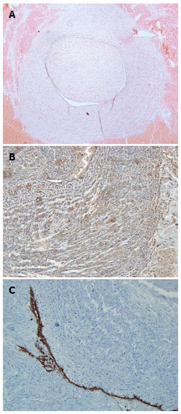
Figure 1 Photographs.
A: Circumscribed intravascular tumor. Micrograph showing the intravascular tumor lined with endothelium (hematoxylin-eosin staining, magnification × 20); B: Smooth muscle actin immunohistochemistry. Micrograph showing tumor cells immunoreactive for smooth muscle actin (magnification × 20); C: CD34+ immunoreactivity in tumor cells. Immunohistochemistry revealed the presence of CD34+ tumor cells beneath the endothelium (magnification × 20).
- Citation: Rovas L, Dauksas R, Simavicius A. Leiomyoma of the umbilical cord artery: A case report. World J Obstet Gynecol 2014; 3(3): 138-140
- URL: https://www.wjgnet.com/2218-6220/full/v3/i3/138.htm
- DOI: https://dx.doi.org/10.5317/wjog.v3.i3.138


