Copyright
©The Author(s) 2015.
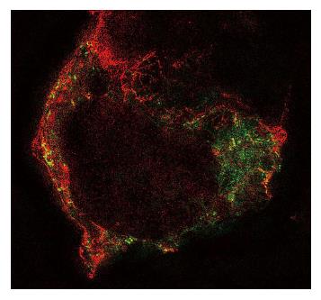
Figure 5 Structured illumination image of an human embryonic kidney cell cotransfected with human PIEZO1 channels labelled green and TREK-1 channels labelled red using green fluorescent protein mutants.
Notice that the channels are in different structural domains and thus feel different forces. Notice also that TREK-1 tends to follow underlying cytoskeletal fibers. (Courtesy Gottlieb P).
- Citation: Sachs F. Mechanical transduction by ion channels: A cautionary tale. World J Neurol 2015; 5(3): 74-87
- URL: https://www.wjgnet.com/2218-6212/full/v5/i3/74.htm
- DOI: https://dx.doi.org/10.5316/wjn.v5.i3.74


