Copyright
©2013 Baishideng Publishing Group Co.
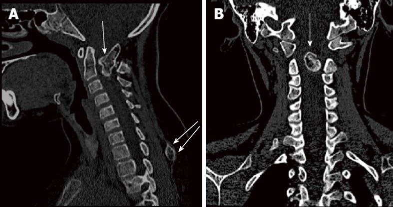
Figure 1 Computed tomography scan of the cervical spine.
A: Sagittal; B: Coronal reconstruction showing a bony outgrowth filling the spinal canal, arising from the inner posterior arch of C1 at the left site, growing anteriorly and obliterating most of the spinal canal (top single arrow). Another exostosis was incidentally found at the spinous process of T1 (bottom double arrows).
- Citation: Elgamal EA. Complete recovery of severe tetraparesis after excision of large C1-osteochondroma. World J Neurol 2013; 3(3): 79-82
- URL: https://www.wjgnet.com/2218-6212/full/v3/i3/79.htm
- DOI: https://dx.doi.org/10.5316/wjn.v3.i3.79


