Copyright
©2013 Baishideng Publishing Group Co.
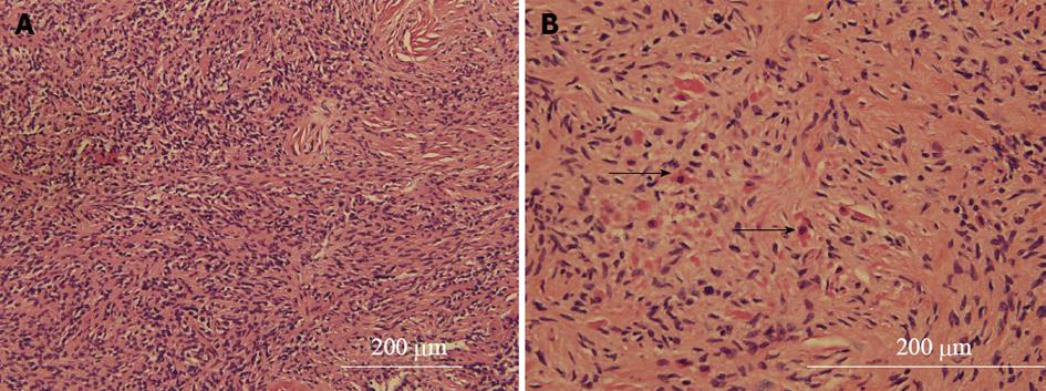
Figure 2 Histopathology of the tumor.
A: High-power micrographs show a spindle cell lesion with focally increased cellularity and nuclear atypia; B: Scattered clusters of mature-appearing, small round cells with moderately abundant eosinophilic cytoplasm (arrow head) resembling a skeletal muscle differentiation.
- Citation: Zhang M, Weaver M, Khurana JS, Mukherjee AL. Low grade spinal malignant triton tumor with mature skeletal muscle differentiation. World J Neurol 2013; 3(3): 75-78
- URL: https://www.wjgnet.com/2218-6212/full/v3/i3/75.htm
- DOI: https://dx.doi.org/10.5316/wjn.v3.i3.75


