Copyright
©2013 Baishideng Publishing Group Co.
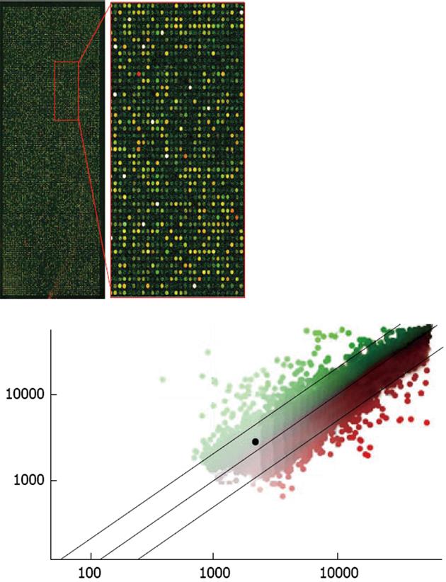
Figure 3 Example of an Agilent Rat Oligo microarray hybridized with probes from microdissected neurons.
The upper, right-hand panel is an enlarged view of a portion of the microarray. The two probes used were made from RNA amplified through one stage of the T7 method, the Cy3-labeled probe was synthesized from total RNA extracted from several thousand CA1 pyramidal neurons and the Cy5-labeled probe was synthesized from several thousand dentate gyrus granule cells, after laser-assisted microdissection. The two probes are shown overlaid; the predominance of yellow spots indicates that most of the genes in the two samples were at or near equivalent levels. Only a few spots are saturated (white). Shown in the lower panel, the genes of interest can be identified on the scatterplot (CA1 neurons vs dentate granular neurons). An example of one gene of interest is the highlighted black spot, which represents caspase-3, a key apoptotic mediator.
- Citation: He Z, Cui L, He B, Ferguson SA, Paule MG. A common genetic mechanism underlying susceptibility to posttraumatic stress disorder. World J Neurol 2013; 3(3): 14-24
- URL: https://www.wjgnet.com/2218-6212/full/v3/i3/14.htm
- DOI: https://dx.doi.org/10.5316/wjn.v3.i3.14


