Copyright
©The Author(s) 2024.
World J Neurol. Dec 31, 2024; 10(1): 98672
Published online Dec 31, 2024. doi: 10.5316/wjn.v10.i1.98672
Published online Dec 31, 2024. doi: 10.5316/wjn.v10.i1.98672
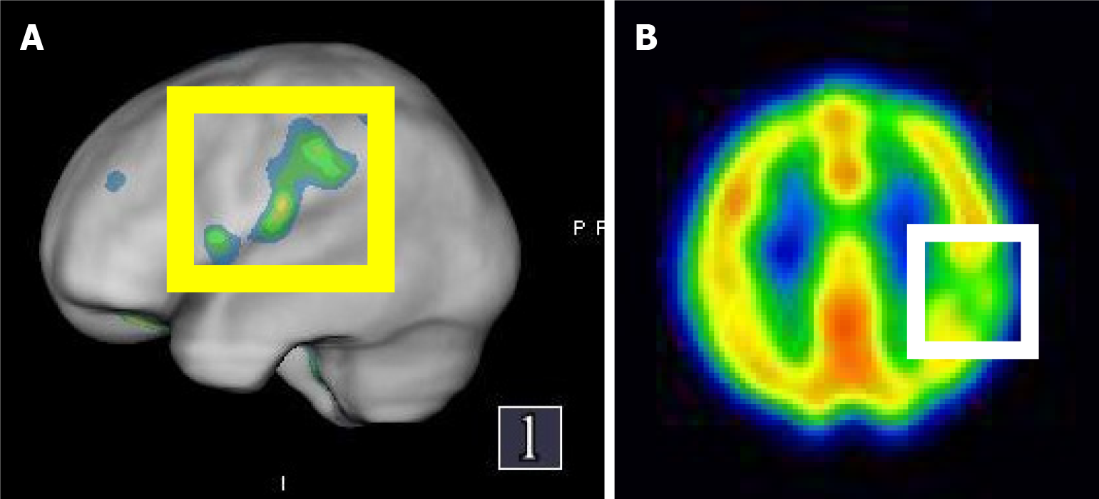
Figure 4 Single-photon emission computed tomography images of the patient.
A: Single-photon emission computed tomography (SPECT) images of the patient’s temporal lobe, showing reduced blood perfusion below the left parietal lobe in the SPECT image with “Q. brain” software; B: Shows decreased uptake in the left parietal lobe of the transverse image SPECT.
- Citation: Tsai YH, Chen YH, Chao TC, Lin LF, Chang ST. New type of lacunar stroke presenting in brain perfusion images: A case report. World J Neurol 2024; 10(1): 98672
- URL: https://www.wjgnet.com/2218-6212/full/v10/i1/98672.htm
- DOI: https://dx.doi.org/10.5316/wjn.v10.i1.98672


