Copyright
©The Author(s) 2024.
World J Neurol. Dec 31, 2024; 10(1): 98672
Published online Dec 31, 2024. doi: 10.5316/wjn.v10.i1.98672
Published online Dec 31, 2024. doi: 10.5316/wjn.v10.i1.98672
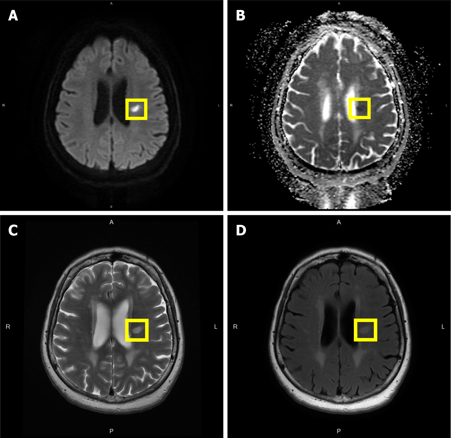
Figure 1 Magnetic resonance images of the patient.
A: Diffusion weighted imaging images, acute lacunar infarct in the left corona radiata; B: Apparent diffusion coefficient images; C: T2 images; D: T2 fluid attenuated inversion recovery images, hyperintense changes in the subcortical and periventricular white matter indicating small vessel ischemic changes; and old lacunar infarcts in bilateral centrum semiovale.
- Citation: Tsai YH, Chen YH, Chao TC, Lin LF, Chang ST. New type of lacunar stroke presenting in brain perfusion images: A case report. World J Neurol 2024; 10(1): 98672
- URL: https://www.wjgnet.com/2218-6212/full/v10/i1/98672.htm
- DOI: https://dx.doi.org/10.5316/wjn.v10.i1.98672


