Copyright
©The Author(s) 2017.
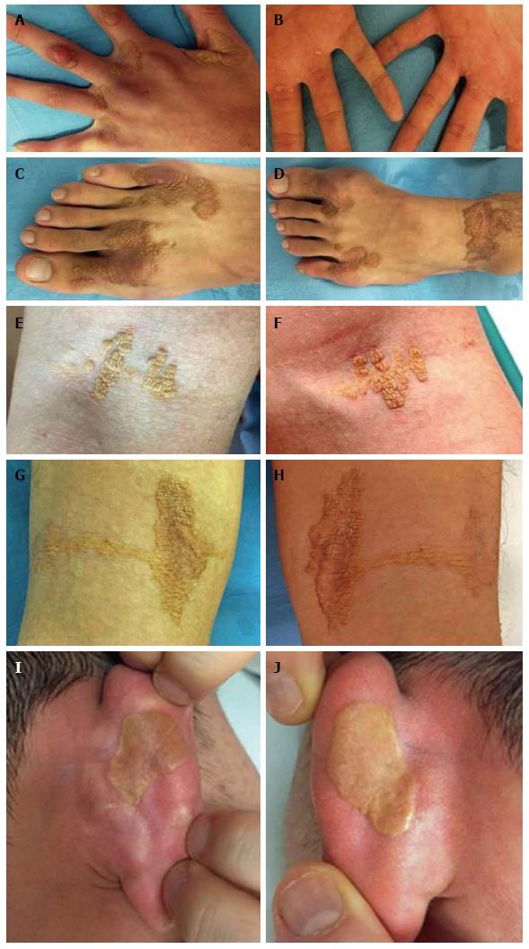
Figure 3 Diffuse intertriginous xanthomas.
Usually appear in a symmetric distribution as well-demarcated and slightly elevated noninflammatory plaques of ochre-yellow or yellow-brown discoloration. Typically found in intertriginous and flexural areas. A: In finger web spaces, and in this picture with metacarpophalangeal joint tendon xanthoma; B: At metacarpophalangeal palmar crease in linear band or single papules; C: Toe web spaces; D: In toe web spaces and ankle crease; E and F: At antecubital fossae, with the “eruptive” appearance of crops of yellow dermal soft, velvety papules; G and H: In popliteal fossae; I and J: At the creases of ears in a rare pattern of “plane xanthoma” as very thin flat patches, easily clinically missed, of yellow-orange macular discoloration.
- Citation: Mastrolorenzo A, D’Errico A, Pierotti P, Vannucchi M, Giannini S, Fossi F. Pleomorphic cutaneous xanthomas disclosing homozygous familial hypercholesterolemia. World J Dermatol 2017; 6(4): 59-65
- URL: https://www.wjgnet.com/2218-6190/full/v6/i4/59.htm
- DOI: https://dx.doi.org/10.5314/wjd.v6.i4.59


