Copyright
©The Author(s) 2015.
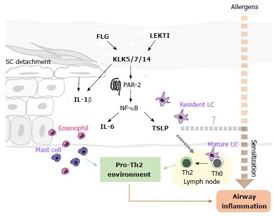
Figure 3 Hypothesised pathways from skin barrier impairment to inflammation and the atopic march.
Deficiencies in FLG and LEKTI promote enhanced activity of KLKs, which induce over-expression of TSLP and IL-6 through the PAR-2-NF-kB pathway. KLK hyperactivity also degrades transition desmosomes, inducing the release of pro-inflammatory cytokines from mechanically stressed keratinocytes. The pro-inflammatory environment triggers eosinophil and mast cell recruitment and activation. TSLP activates LCs which promote the differentiation of naïve (Th0) T cells into Th2 cells in lymph nodes. Allergen ingress (dashed red arrow) promotes allergic sensitization and may, with TSLP, stimulate downstream airway inflammation. FLG: Filaggrin; LEKTI: Lympho-epithelial Kazal-type-related inhibitor; KLKs: Kallikrein proteases; TSLP: Thymic stromal lymphopoietin; IL-6: Interleukin-6; IL-1β: Interleukin-1β; LCs: Langerhans cells; SC: Stratum corneum; PAR-2: Protease-activated receptor-2; NF-kB: Nuclear factor kappa-light-chain-enhancer of activated B cells.
- Citation: Gillespie RM, Brown SJ. From the outside-in: Epidermal targeting as a paradigm for atopic disease therapy. World J Dermatol 2015; 4(1): 16-32
- URL: https://www.wjgnet.com/2218-6190/full/v4/i1/16.htm
- DOI: https://dx.doi.org/10.5314/wjd.v4.i1.16


