Copyright
©2014 Baishideng Publishing Group Inc.
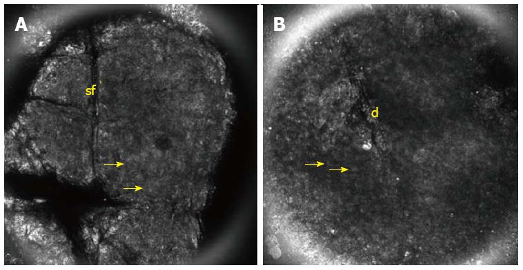
Figure 1 Reflectance confocal microscopy image (0.
5 mm × 0.5 mm). A: Shows preservation of the stratum corneum in early stages of allergic dermatitis with parakeratotic corneocytes (yellow arrow) between skin folds (sf); B: Shows disruption (d) with early parakeratosis of the stratum corneum in irritative contact dermatitis.
- Citation: Suárez-Pérez JA, Bosch R, González S, González E. Pathogenesis and diagnosis of contact dermatitis: Applications of reflectance confocal microscopy. World J Dermatol 2014; 3(3): 45-49
- URL: https://www.wjgnet.com/2218-6190/full/v3/i3/45.htm
- DOI: https://dx.doi.org/10.5314/wjd.v3.i3.45


