Copyright
©The Author(s) 2015.
World J Anesthesiol. Jul 27, 2015; 4(2): 30-38
Published online Jul 27, 2015. doi: 10.5313/wja.v4.i2.30
Published online Jul 27, 2015. doi: 10.5313/wja.v4.i2.30
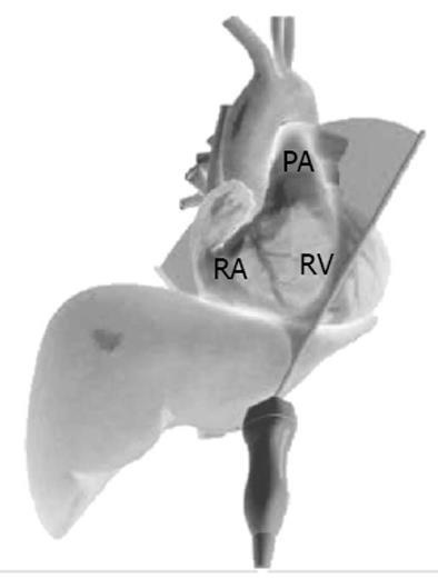
Figure 4 Subcostal right ventricular inflow outflow view, antero-inferior projection.
Schematic diagram demonstrating TTE probe position and alignment of scanning plane. Reproduced in part with permission from Toronto General Hospital, Perioperative Interactive Education Virtual TTE (http://pie.med.utoronto.ca/TTE). TTE: Transthoracic echocardiogram; RA: Right atrium; RV: Right ventricle; PA: Pulmonary artery.
- Citation: Tan CO, Weinberg L, Story DA, McNicol L. Transthoracic echocardiography assists appropriate pulmonary artery catheter placement: An observational study. World J Anesthesiol 2015; 4(2): 30-38
- URL: https://www.wjgnet.com/2218-6182/full/v4/i2/30.htm
- DOI: https://dx.doi.org/10.5313/wja.v4.i2.30


