Copyright
©The Author(s) 2018.
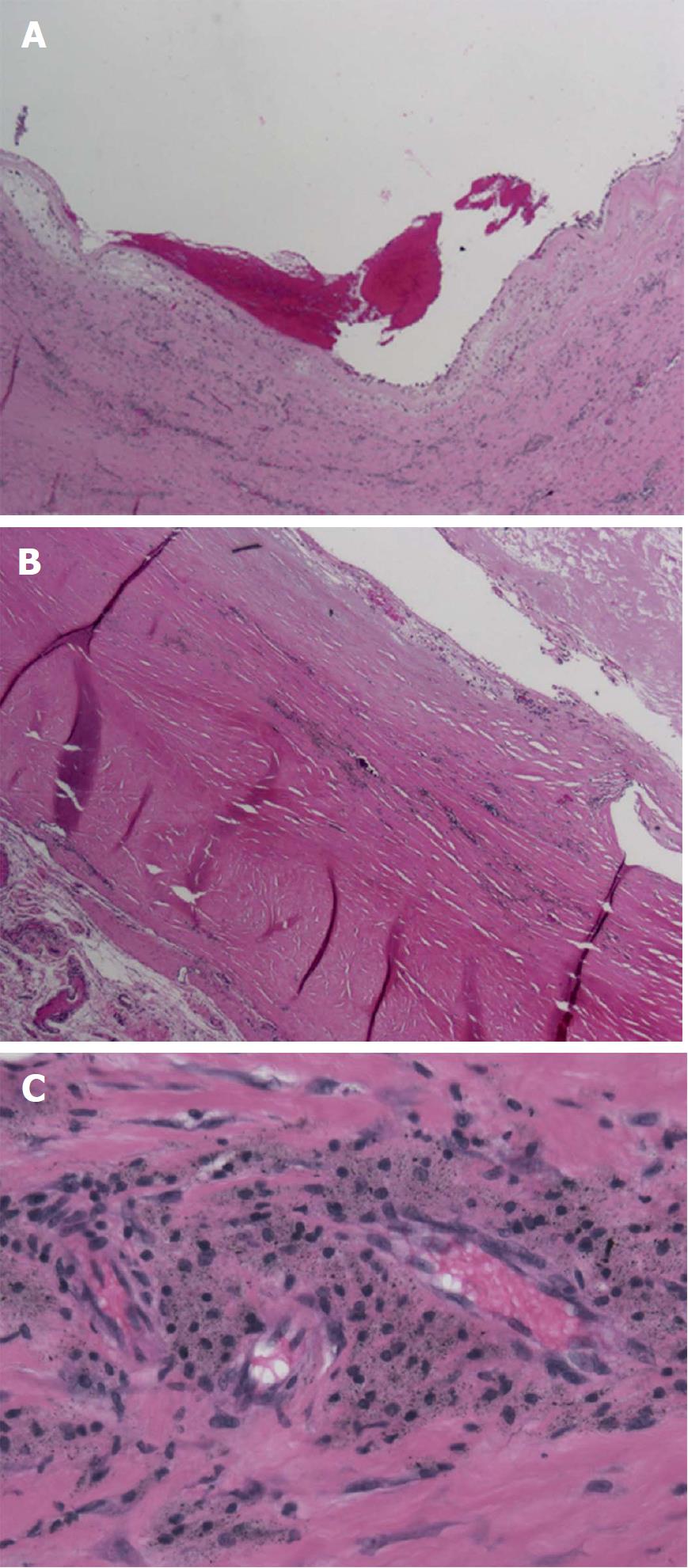
Figure 5 Histologic examination of the pseudotumor with specimen taken in the middle of the cystic mass.
A: Low power (100 ×) view of mass demonstrating its cystic nature with blood and fibrin products within the centre of cystic structure. The wall of the lesion is very thick, studded with islands of metallic particles; B: Intermediate power (200 ×) view of the wall of pseudotumor mass. In the lower left corner, the cellular reaction is primarily lymphocytic and histiocytic with perivascular lymphocytes. The eosinophilic (red-color) area shows the pseudotumor cyst wall with adherent fibrin debris (top right corner) and microscopic metal particles within the wall; C: High power (400 ×) view of the cellular reaction surrounding pseudotumor mass. This shows the "classic" histologic finding of a reactive pseudotumor due to small metallic particles. The micrograph shows perivascular infiltration of histiocytes, containing particles of metallic debris, along with lymphocytes.
- Citation: Chowdhry M, Dipane MV, McPherson EJ. Periosteal pseudotumor in complex total knee arthroplasty resembling a neoplastic process. World J Orthop 2018; 9(5): 72-77
- URL: https://www.wjgnet.com/2218-5836/full/v9/i5/72.htm
- DOI: https://dx.doi.org/10.5312/wjo.v9.i5.72


