Copyright
©The Author(s) 2018.
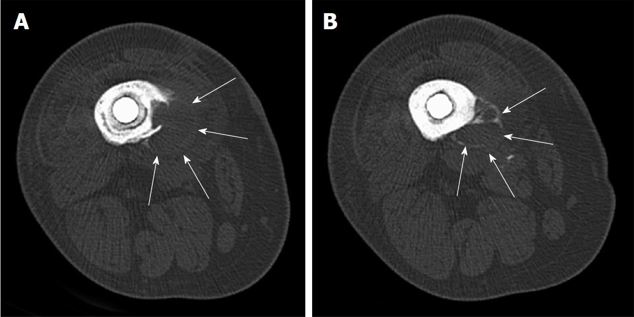
Figure 2 Transverse computed tomography scan images of distal femoral diaphysis centered over lytic lesion.
A: Image shows a surrounding soft tissue mass of 2.8 cm. Erosive lytic lesion extends down to the inner cortex. Also note the radiolucent lines between the prosthetic implant and cement mantle, as well as the radiolucent line between the cement mantle and bone; B: Demonstrates septation of the soft tissue mass with peripheral calcification (arrows). This is typically a worrisome sign for a neoplastic process.
- Citation: Chowdhry M, Dipane MV, McPherson EJ. Periosteal pseudotumor in complex total knee arthroplasty resembling a neoplastic process. World J Orthop 2018; 9(5): 72-77
- URL: https://www.wjgnet.com/2218-5836/full/v9/i5/72.htm
- DOI: https://dx.doi.org/10.5312/wjo.v9.i5.72


