Copyright
©The Author(s) 2018.
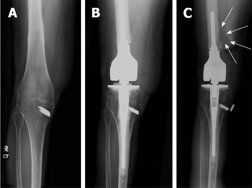
Figure 1 AP radiograph of right knee reviewing life-cycle of right knee reconstruction.
A: Pre-operative radiograph showing successful knee fusion. The area over the medial staple has a split thickness skin graft (STSG) of 5 cm x 7 cm. Patient ambulates with full weight bearing on the right leg. Localized osteopenia is present in the knee region; B: Four months post-operative radiograph of cemented endoprosthetic hinge total knee arthroplasty using a modified Zimmer Biomet RS (Reduced Size) OSS Knee system. Note particularly the placement of the joint line in the cephalad direction. This was purposefully done to avoid dissecting near the area of the tibia with the STSG. Also, note the relatively short cemented femoral stem; C: Radiograph at 6 years. Patient complains of mild pain for the past 5 months. Note the erosive lesion in the distal femoral diaphysis. White arrows outline the soft tissue mass surrounding the erosive lesion. Also note the radiolucent line in the distal femoral diaphysis. This suggests cement-bone separation, likely from repetitive cantilever bend.
- Citation: Chowdhry M, Dipane MV, McPherson EJ. Periosteal pseudotumor in complex total knee arthroplasty resembling a neoplastic process. World J Orthop 2018; 9(5): 72-77
- URL: https://www.wjgnet.com/2218-5836/full/v9/i5/72.htm
- DOI: https://dx.doi.org/10.5312/wjo.v9.i5.72


