Copyright
©The Author(s) 2017.
World J Orthop. Sep 18, 2017; 8(9): 735-740
Published online Sep 18, 2017. doi: 10.5312/wjo.v8.i9.735
Published online Sep 18, 2017. doi: 10.5312/wjo.v8.i9.735
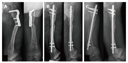
Figure 3 Clinical presentation of case 3 during the first operation.
A 16-year-old male with type III osteogenesis imperfecta presented with subtrochanteric peri-implant fracture of left femur after corrective osteotomy and fixation with osteotomy plate for 10 mo. Plain radiographs in anteroposterior and lateral views after injury (A), immediately after humeral nail fixation (B), and 8 mo after humeral nail fixation (C).
- Citation: Sa-ngasoongsong P, Saisongcroh T, Angsanuntsukh C, Woratanarat P, Mulpruek P. Using humeral nail for surgical reconstruction of femur in adolescents with osteogenesis imperfecta. World J Orthop 2017; 8(9): 735-740
- URL: https://www.wjgnet.com/2218-5836/full/v8/i9/735.htm
- DOI: https://dx.doi.org/10.5312/wjo.v8.i9.735


